腸胃道與腹壁超音波
01使用時機與本章範圍
急性腹痛的整體掃描策略、游離氣體與腸阻塞,已在 第 10 章 急性腹痛 介紹。本章把鏡頭拉近到腸道本身:怎麼認出腸段、怎麼判讀腸壁,以及急診最常遇到的幾個腸胃道與腹壁診斷。
學完這一章,你應該能夠
- 說出正常腸壁的五層構造,並從皺襞、結腸袋與位置分辨胃、小腸與大腸。
- 用漸進加壓(graded compression)找闌尾,說出闌尾炎的直接與間接徵象。
- 認出腸套疊的標靶徵象與假腎徵象,以及憩室炎、疝氣嵌頓的警訊。
- 用胃竇評估胃內容物,並知道超音波確認鼻胃管位置的限制。
什麼時候拿探頭
- 右下腹痛:成人與兒童懷疑闌尾炎,尤其想避免輻射的兒童與孕婦。
- 幼兒陣發性哭鬧、腹痛或嘔吐:懷疑腸套疊。
- 左下腹痛:懷疑憩室炎;右下腹痛也要想到盲腸憩室炎。
- 腹壁或鼠蹊腫塊:疝氣、疝氣嵌頓、腹壁膿瘍。
- 插管或鎮靜前:胃裡還有沒有東西(本課程講義)。
本章的敏感度、特異度只引用已發表的原始研究或統合分析;講義中的原則以「本課程講義」標示。
02腸道的超音波解剖與掃描技巧
腸壁在高頻探頭下呈現亮暗相間的「腸壁特徵」(gut signature)。一旦認得這五層,就能分辨眼前的管狀構造是腸道、血管還是其他東西,並判斷腸壁是否增厚、分層是否還在。
五層構造(由腔內往外,本課程講義)
| 層 | 回音 | 對應組織 |
|---|---|---|
| 1 | 高回音 | 腔內內容物與黏膜的交界面 |
| 2 | 低回音 | 黏膜肌層(muscularis mucosa) |
| 3 | 高回音 | 黏膜下層(submucosa) |
| 4 | 低回音 | 固有肌層(muscularis propria) |
| 5 | 高回音 | 漿膜(serosa) |
怎麼認腸段
- 胃:肌層較厚、黏膜有皺褶(rugae);胃竇在肝左葉後方、胰臟前方。
- 空腸:環狀皺襞(circular folds / Kerckring folds)明顯,常位於左上腹。
- 迴腸:皺襞較少,往右下腹接到盲腸。
- 大腸:有結腸袋(haustra)、常含氣體與糞便;升、降結腸與直腸在腹膜後,位置固定,橫結腸與乙狀結腸在腹膜內、會移動(本課程講義)。
探頭與手法(本課程講義)
- 凸面探頭(約 2.5–5 MHz):穿透較深,先全腹掃描找擴張腸道、積液與腫塊。
- 線性探頭(約 7–15 MHz):軸向解析度較好,看腸壁分層、闌尾;體型瘦或兒童特別好用。
- 輕柔、穩定、持續地加壓,把腸氣推開;壓痛最明顯的地方往往就是病灶。
- 看胃時:平躺加右側臥;需要時請病人深吸氣、左側臥,或喝水把胃撐開當聲窗。
系統性掃全腹
- 從右上腹橫放凸面探頭,往下掃到右下腹,再往內側移 1–2 cm 一路掃回來,像割草一樣涵蓋全腹(Theophanous 2026)。
- 也可以直接從壓痛最明顯處開始,再擴大範圍。
- 每個異常腸段都記錄:位置、管徑、壁厚與分層、蠕動、周圍脂肪與積液、都卜勒血流。
正常小腸管徑一般 < 3 cm、大腸 < 6 cm(Theophanous 2026);腸阻塞的判讀詳見 急性腹痛章。
03腸壁增厚:看厚度、分層、範圍與血流
腸壁增厚本身不是診斷,而是一個要繼續追問的發現。結腸壁正常上限約 2–3 mm(Boccatonda 2025 圖例);克隆氏症常以 > 3 mm 為異常(敏感度 89%、特異度 96%,Boccatonda 2025 引述)。接著問四件事:多厚、分層還在不在、侵犯多長、血流多或少。
| 型態 | 先想到 | 重點 |
|---|---|---|
| 整段大腸連續、黏膜與黏膜下層增厚、結腸袋消失 | 潰瘍性結腸炎 | 本課程講義 |
| 跳躍式、全層侵犯,黏膜下層特別厚;可有膿瘍、瘻管、腸沾黏成團 | 克隆氏症 | 本課程講義 |
| 皺襞輕度水腫、蠕動過強、黏膜下層增厚 | 感染性腸炎 | 本課程講義 |
| 分段環狀低回音增厚、結腸旁脂肪變亮、血流減少或消失、與正常腸段交界突然 | 缺血性大腸炎 | Boccatonda 2025 |
| 局部不規則增厚、分層消失、不可壓(假腎外觀) | 腫瘤 | Chen 2022 病例 |
表格為判讀方向,不是診斷標準;確診仍靠內視鏡、CT 或病理。
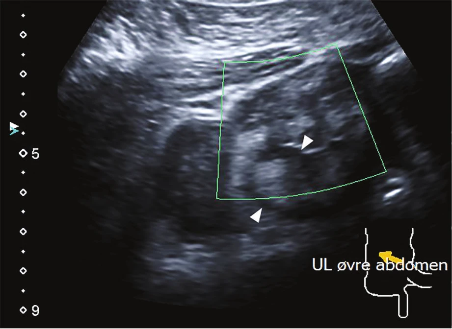
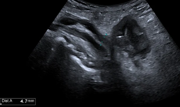
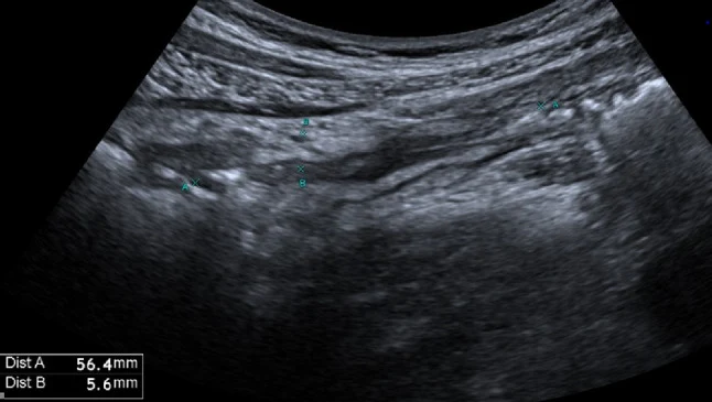
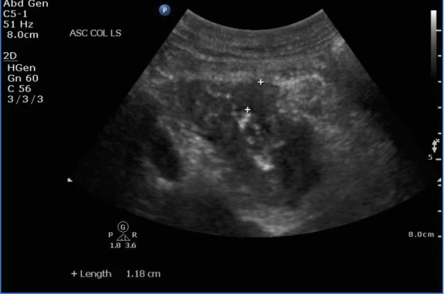
影像來源:Leth 2015(Case Rep Gastrointest Med)、Chen 2022(POCUS J)、Boccatonda 2025(J Ultrasound),CC BY 4.0;Fig. 8–9 原文圖說相同,此處分述量測重點。
04闌尾炎(成人與兒童)
在壓痛最明顯處用線性探頭漸進加壓,把腸氣慢慢推開。先找到盲腸與迴腸末端,再在腰大肌與髂血管前方找一條盲端、不蠕動的管狀構造。正常闌尾可以壓扁;講義以加壓下外徑 < 6 mm、可壓扁為正常。
直接徵象
- 盲端、不可壓的管狀構造,外徑 > 6 mm(Boccatonda 2025)。
- 短軸呈標靶狀,壁增厚;探頭壓在上面有壓痛。
- 闌尾糞石:強回音+後方聲影。
- 早期、未穿孔時壁內血流增加。
間接徵象
- 周圍脂肪變亮、不可壓(發炎的腸繫膜脂肪包住闌尾)。
- 闌尾周圍積液或膿瘍、腸繫膜淋巴結腫大。
- 盲腸後位闌尾不易用加壓看到,是偽陰性的常見原因(Boccatonda 2025)。
準確度
- 所有年齡(21 篇統合分析):非放射科醫師的 POCUS 敏感度 91%、特異度 97%;急診醫師操作的研究較低(80%/92%),研究間異質性很大(Fields 2017)。
- 兒童急診(21 篇、8,605 人):急診 POCUS 陽性概似比 9.24、陰性概似比 0.17;陽性可直接確立,陰性不足以排除(Benabbas 2017)。
- 結論都一樣:陽性有用,陰性或看不到闌尾時不能排除,依臨床機率再掃、觀察或做 CT/MRI。
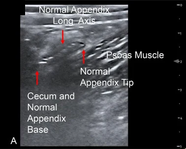
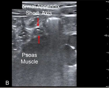
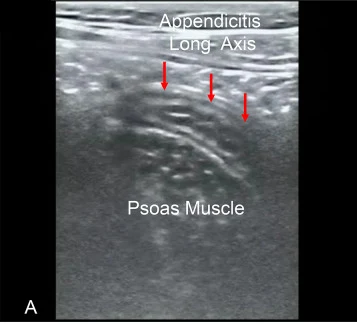
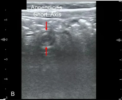
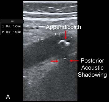
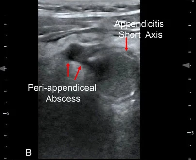
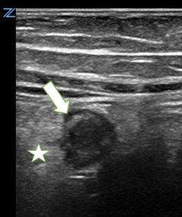
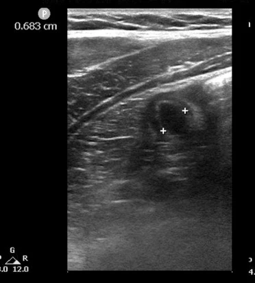
影像來源:Tsung 2024(Ultrasound J)、Scheier 2024、Kaplan 2025(POCUS J),裁切單格,CC BY 4.0;Tsung 2024 影像角落的機型標誌已遮蓋。
05腸套疊
一段腸子(套入部)連同它的腸繫膜,套進遠端的腸子裡。兒童多為迴腸–結腸型、多半沒有病理性前導點;成人的腸套疊則常有前導點(腫瘤等),要找原因(Theophanous 2026)。
怎麼掃
- 用線性探頭(小孩)或凸面探頭(大人),從右上腹開始:迴腸–結腸型最常見(Theophanous 2026)。
- 沿著結腸走一圈,像畫框一樣把腹部周邊掃完(picture-frame);也可以直接從壓痛最明顯處找起。
- 短軸找標靶(target/doughnut)徵象;長軸找假腎(pseudokidney)或三明治徵象。
- 看到之後:量直徑、看是否有前導點、周圍積液、腸壁血流,以及上游腸道是否擴張。
準確度
- 兒童急診 POCUS(6 篇、1,303 人):敏感度 94.9%、特異度 99.1%、陽性概似比 105、陰性概似比 0.05(Lin-Martore 2020)。
- 學得快:小兒急診醫師只接受 1 小時訓練,82 位病童中敏感度 85%、特異度 97%(Riera 2012)。
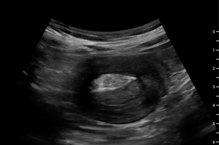
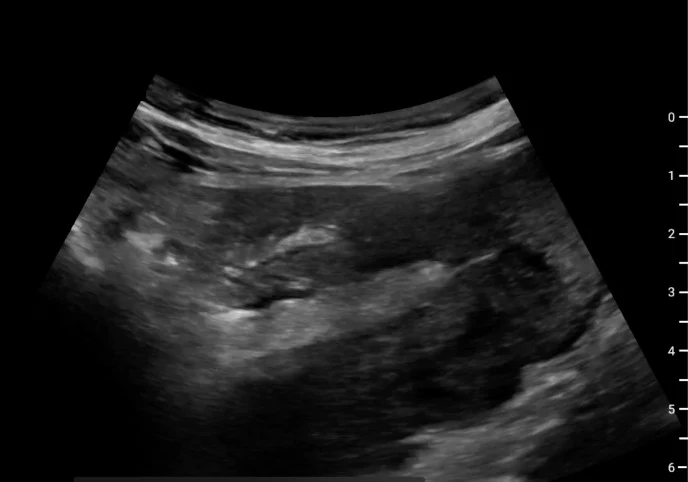
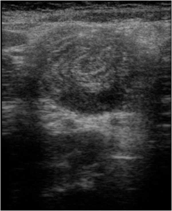
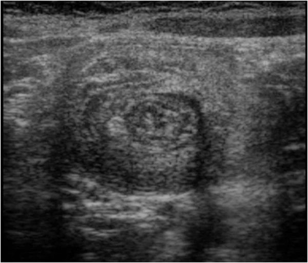
影像來源:Greenwald 2025(POCUS J,CC BY 4.0,探頭標誌已遮蓋)、Fonio 2013(Crit Ultrasound J,CC BY 2.0)。
以下內容需要免費訂閱
病例影像判讀與練習題開放給訂閱者。留下電子郵件即可立即閱讀全部內容;同一個瀏覽器一年內不必再輸入。換手機或電腦時,輸入同一個信箱即可。
不收費、不寄廣告。若想收到新章節通知,我們會另寄一封確認信,點了才會開始寄。詳見隱私說明。
12參考文獻與延伸閱讀
- Nylund K, Maconi G, Hollerweger A, et al. EFSUMB recommendations and guidelines for gastrointestinal ultrasound. Ultraschall Med. 2017;38(3):273-284. doi:10.1055/s-0042-115410
- Hollerweger A, Maconi G, Ripolles T, et al. Gastrointestinal ultrasound (GIUS) in intestinal emergencies - an EFSUMB position paper. Ultraschall Med. 2020;41(6):646-657. doi:10.1055/a-1147-1295
- Boccatonda A, Brighenti A, Tiraferri V, et al. POCUS for acute abdominal pain: practical scan protocols on gastrointestinal diseases and an evidence review. J Ultrasound. 2025;28(4):851-871. doi:10.1007/s40477-025-01088-7
- Theophanous RG, Abramson L, Bronshteyn YS. Identifying gastrointestinal pathologies using point-of-care ultrasound. Diagnostics (Basel). 2026;16(3):418. doi:10.3390/diagnostics16030418
- Fields JM, Davis J, Alsup C, et al. Accuracy of point-of-care ultrasonography for diagnosing acute appendicitis: a systematic review and meta-analysis. Acad Emerg Med. 2017;24(9):1124-1136. doi:10.1111/acem.13212
- Benabbas R, Hanna M, Shah J, Sinert R. Diagnostic accuracy of history, physical examination, laboratory tests, and point-of-care ultrasound for pediatric acute appendicitis in the emergency department: a systematic review and meta-analysis. Acad Emerg Med. 2017;24(5):523-551. doi:10.1111/acem.13181
- Lin-Martore M, Kornblith AE, Kohn MA, Gottlieb M. Diagnostic accuracy of point-of-care ultrasound for intussusception in children presenting to the emergency department: a systematic review and meta-analysis. West J Emerg Med. 2020;21(4):1008-1016. doi:10.5811/westjem.2020.4.46241
- Riera A, Hsiao AL, Langhan ML, Goodman TR, Chen L. Diagnosis of intussusception by physician novice sonographers in the emergency department. Ann Emerg Med. 2012;60(3):264-268. doi:10.1016/j.annemergmed.2012.02.007
- Zhang W, Sun H, Luo F. The efficiency of sonography in diagnosing volvulus in neonates with suspected intestinal malrotation. Medicine (Baltimore). 2017;96(42):e8287. doi:10.1097/MD.0000000000008287
- Laméris W, van Randen A, Bipat S, et al. Graded compression ultrasonography and computed tomography in acute colonic diverticulitis: meta-analysis of test accuracy. Eur Radiol. 2008;18(11):2498-2511. doi:10.1007/s00330-008-1018-6
- Robinson A, Light D, Nice C. Meta-analysis of sonography in the diagnosis of inguinal hernias. J Ultrasound Med. 2013;32(2):339-346. doi:10.7863/jum.2013.32.2.339
- Kwee RM, Kwee TC. Ultrasonography in diagnosing clinically occult groin hernia: systematic review and meta-analysis. Eur Radiol. 2018;28(11):4550-4560. doi:10.1007/s00330-018-5489-9
- Hosokawa T, Yoshizawa S, Deie K, et al. Point-of-care ultrasound for mimicker lesions of incarcerated inguinal hernia. Case Rep Pediatr. 2023;2023:5593369. doi:10.1155/2023/5593369
- Perlas A, Davis L, Khan M, Mitsakakis N, Chan VW. Gastric sonography in the fasted surgical patient: a prospective descriptive study. Anesth Analg. 2011;113(1):93-97. doi:10.1213/ANE.0b013e31821b98c0
- Perlas A, Mitsakakis N, Liu L, et al. Validation of a mathematical model for ultrasound assessment of gastric volume by gastroscopic examination. Anesth Analg. 2013;116(2):357-363. doi:10.1213/ANE.0b013e318274fc19
- Kim HM, So BH, Jeong WJ, Choi SM, Park KN. The effectiveness of ultrasonography in verifying the placement of a nasogastric tube in patients with low consciousness at an emergency center. Scand J Trauma Resusc Emerg Med. 2012;20:38. doi:10.1186/1757-7241-20-38
- Marron A, Wolf MS, Levine M, et al. The utilization of point of care ultrasound (POCUS) for the confirmation of gastric and post-pyloric feeding tube placement in a pediatric intensive care unit. POCUS J. 2025;10(1):164-174. doi:10.24908/pocusj.v10i01.17785
- Novak KL, Jacob D, Kaplan GG, et al. Point of care ultrasound accurately distinguishes inflammatory from noninflammatory disease in patients presenting with abdominal pain and diarrhea. Can J Gastroenterol Hepatol. 2016;2016:4023065. doi:10.1155/2016/4023065
圖片來源與授權
- 以下論文的多格拼圖在本頁拆成單格裁切呈現,圖說為本站中文改寫;影像上的機型或探頭標誌、檢查編號與原圖上方的文字列已遮蓋或裁除。
- Tsung JW, Firnberg M, Sosa P. Interobserver agreement of an ED PoCUS video training dataset of normal appendix and appendicitis in children. Ultrasound J 2024;16:38, doi:10.1186/s13089-024-00386-1(Fig. 1–3),CC BY 4.0。
- Scheier E, Taragin B. Resolution of sonographic appendicitis in pediatrics: a point of care ultrasound case-series. POCUS J 2024;9(1):44-50, doi:10.24908/pocus.v9i1.16860(Fig. 2),CC BY 4.0。
- Kaplan C, Vaid R, Secko M. Identification of pediatric retrocecal appendicitis using point of care ultrasound (POCUS). POCUS J 2025;10(1):192-195, doi:10.24908/pocusj.v10i01.17744(Fig. 2),CC BY 4.0。
- Greenwald S, Ramos M, Kohen B. Man with abdominal bloating, weight loss. POCUS J 2025;10(1):123-124, doi:10.24908/pocusj.v10i01.18109(Fig. 1、2),CC BY 4.0。
- Fonio P, Coppolino F, Russo A, et al. Ultrasonography (US) in the assessment of pediatric non traumatic gastrointestinal emergencies. Crit Ultrasound J 2013;5(Suppl 1):S12, doi:10.1186/2036-7902-5-S1-S12(Fig. 1、2),CC BY 2.0。
- Mazzei MA, Cioffi Squitieri N, Guerrini S, et al. Sigmoid diverticulitis: US findings. Crit Ultrasound J 2013;5(Suppl 1):S5, doi:10.1186/2036-7902-5-S1-S5(Fig. 2),CC BY 2.0。
- Theophanous RG, Abramson L, Bronshteyn YS. Identifying gastrointestinal pathologies using point-of-care ultrasound. Diagnostics (Basel) 2026;16(3):418, doi:10.3390/diagnostics16030418(Fig. 2、3),CC BY 4.0。
- Boccatonda A, Brighenti A, Tiraferri V, et al. POCUS for acute abdominal pain: practical scan protocols on gastrointestinal diseases and an evidence review. J Ultrasound 2025;28(4):851-871, doi:10.1007/s40477-025-01088-7(Fig. 6、8、9),CC BY 4.0。
- Chen W, Teh R, Qurishi A. Point-of-care ultrasound for the diagnosis of colon cancer. POCUS J 2022;7(2):190-192, doi:10.24908/pocus.v7i2.15657(Fig. 1),CC BY 4.0。
- Lochner A, Stovicek E, Kman N, et al. Ultrasound-guided hernia reduction—preventing surgery for an incarcerated ventral hernia: a case report. Clin Pract Cases Emerg Med 2026;10(3):354-357, doi:10.5811/cpcem.47051(Image 1、2),CC BY 4.0。
- Desai N, Lawson J, Elwen F, Howle R. The pregnant and postpartum stomach. Eur J Anaesthesiol 2026;43(2):103-118, doi:10.1097/EJA.0000000000002320(Fig. 2–4),CC BY 4.0。
- Koveleskie J, Labat S, Palascak M, et al. Benefits of point-of-care ultrasound for assessing gastric contents. Ochsner J 2026;26(3):262-265, doi:10.31486/toj.26.0002(Fig. 3),CC BY 4.0。
- McCuskee S, Anderson KL. Enterocutaneous fistula and abscess diagnosed with point-of-care ultrasound. Clin Pract Cases Emerg Med 2021;5(4):470-472, doi:10.5811/cpcem.2021.4.49918(Image 1),CC BY 4.0。
- Uezu I, Takamiyagi Y, Chibana T, et al. Laparoscopic removal of a fish bone causing an abdominal wall abscess using laparoscopic ultrasonography: a case report. Surg Case Rep 2026;12(1), doi:10.70352/scrj.cr.26-0118(Fig. 1A),CC BY 4.0。
- Leth T, Wilkens R, Bonderup OK. Sonographic and endoscopic findings in cocaine-induced ischemic colitis. Case Rep Gastrointest Med 2015;2015:680937, doi:10.1155/2015/680937(Fig. 1a),CC BY 4.0。