急性腹痛的超音波評估
01使用時機:急性腹痛為什麼先拿探頭
急性腹痛是急診最常見的主訴之一,病因從腸胃炎到主動脈瘤破裂都有。床邊超音波(POCUS)的角色不是取代電腦斷層(CT),而是在病人床邊先抓出有時間壓力的急症,再決定誰需要 CT、誰可以直接會診或處置。
學完這一章,你應該能夠
- 用 ACUTE ABDOMEN 口訣列出急性腹痛要用超音波找的 11 類診斷(本課程講義)。
- 依血行動力學與疼痛位置決定先掃哪裡。
- 認出游離氣體(腹膜腔積氣)的超音波徵象,並分辨正常腸氣與假象。
- 說出超音波與 CT 的取捨:什麼時候超音波可以先做、什麼時候還是要 CT。
為什麼值得先掃
- 時間敏感的急症(本課程講義):主動脈瘤破裂、子宮外孕破裂、腸穿孔、卵巢扭轉,晚一步結果差很多。
- 依族群想病因(本課程講義):例如育齡女性先驗孕、想婦產科急症,年長者別漏了主動脈。
- 整合式評估:一支探頭同時看腹腔、骨盆、主動脈、腎臟與膽囊,不必一次只想一個器官。
- 可重複:腸阻塞、腹水量可以隨時再掃,追蹤變化。
本章是急性腹痛的整體架構。各器官的完整掃描與判讀,請見後續的主動脈、肝膽、腎泌尿與腸胃道各章。
02ACUTE ABDOMEN:11 類要找的診斷
作者講義把急性腹痛要用超音波找的診斷,排成 A-C-U-T-E A-B-D-O-Me-N 的口訣。前五個字母多是單一器官的診斷,後半段則包含游離氣體、腸阻塞、血管與少見但致命的病因。
| 字母 | 診斷 | 超音波上要找什麼 | 超音波的敏感度/特異度 | 研究與執行者 |
|---|---|---|---|---|
| A | 闌尾炎(Appendicitis) | 管徑變粗、壓不扁的盲端管狀構造;周圍脂肪發炎、積液、糞石 | 84%/91% 兒童 95%/95% | Lee 2019:統合分析 17 篇、2,385 人,急診醫師 POCUS |
| C | 膽囊炎/膽結石(Cholecystitis) | 結石+後方聲影、膽囊壁增厚、超音波 Murphy 徵象;總膽管結石 | 膽囊炎 70.9%/94.4% 膽結石 89.8%/88.0% 總膽管結石 73%/91%(非 POCUS) | 膽囊炎:Wilson 2024,統合分析 7 篇、1,772 人,急診醫師 POCUS 膽結石:Ross 2011,系統性回顧 8 篇、710 人,急診醫師 總膽管結石:Gurusamy 2015,Cochrane 回顧 5 篇、523 人,一般超音波,原文評為研究品質差 |
| U | 尿路結石(Urolithiasis) | 腎水腫、輸尿管擴張、結石本身、閃爍假影(twinkling)、單側輸尿管噴流消失 | 70.2%/75.4%(任何程度腎水腫算陽性) 中度以上腎水腫:特異度 94.4% | Wong 2018:統合分析 5 篇、1,773 人,急診 POCUS |
| T | 扭轉(Torsion) | 卵巢腫大水腫、附屬腫塊或囊腫、血管蒂扭轉(漩渦徵象) | 附屬器扭轉 79%/76%(非 POCUS) | Wattar 2021:統合分析 12 篇、1,187 人,超音波(摘要未載明執行者,非急診 POCUS 專屬) |
| E | 子宮外孕(Ectopic pregnancy) | 子宮內看不到妊娠、附屬器腫塊、骨盆腔游離液體 | 敏感度 99.3%(以「看不到子宮內孕」為陽性) 特異度:摘要未報告 | Stein 2010:統合分析 10 篇、2,057 人,急診醫師 |
| A | 游離氣體(Air, free) | 腹膜線增強(EPSS)合併後方迴響假影 | 95.5%/81.8%(前導研究) 92%/53% EPSS 單一徵象 100%/99% | Nazerian 2015:22 位急診腹痛病人的影片,資深醫師判讀 Chen 2002:188 位疑似中空臟器穿孔,單一研究(摘要未載明執行者) Asrani 2007:600 位連續急性腹痛病人,單一放射科醫師 |
| B | 腸阻塞(Bowel obstruction) | 小腸擴張、蠕動異常(來回擺動或不動)、遠端大腸塌陷 | 92.4%/96.6% | Gottlieb 2018:統合分析 11 篇、1,178 人,超音波(摘要未載明執行者) |
| D | 主動脈瘤/剝離(Dissection/AAA) | 主動脈外徑 ≥ 3 cm;內膜瓣、壁內血腫 | AAA 99%/98% 剝離 93.2%/90.9%(心臟+腹主動脈的 POCUS 流程) | AAA:Rubano 2013,系統性回顧 7 篇、655 人,急診床邊超音波 剝離:Gibbons 2024,多中心前瞻研究、1,314 次 POCUS(44 例剝離);不是只掃腹主動脈的數字 |
| O | 卵巢過度刺激症候群(OHSS) | 試管療程後腹痛:卵巢腫大、腹水 | 缺乏資料 | 急診 POCUS 僅個案報告,查無診斷準確度研究 |
| Me | 腸繫膜疾病(Mesenteric disease) | SMA 症候群(Wilkie 症候群)、SMV 血栓、SMA 剝離 | SMA 症候群:缺乏資料 SMV 血栓:缺乏資料 SMA 剝離:缺乏資料 | 三者都只查到個案報告或小型描述性研究,沒有可靠的超音波敏感度/特異度 |
| N | 腎盂腎炎(Nephritis,APN) | 腎臟腫大、皮髓質分界消失、低回音區、膿瘍或腎內氣體 | POCUS:缺乏資料 顯影劑超音波 98%/100%(非 POCUS) | Mitterberger 2008:單一研究 100 人,顯影劑超音波對照顯影 CT(摘要未載明執行者) |
口訣與各項目來自作者講義;AAA 定義依 Rubano 2013。數字為「敏感度/特異度」,都取自各研究的 PubMed 摘要。各研究的族群、執行者與參考標準不同,數字不能直接橫向比較;標示「非 POCUS」的是一般超音波或顯影劑超音波的研究,不能當成床邊超音波的表現。「缺乏資料」表示查不到可靠的超音波診斷準確度研究,不代表超音波沒有用。各器官細節見腸胃道、肝膽、腎泌尿、主動脈與婦產相關章節;游離氣體、OHSS 與腸繫膜疾病的影像見本章。
03整合式掃描策略:先看血壓,再看位置
血行動力學不穩定
- 先找出血:右上腹、左上腹、骨盆腔有沒有游離液體。
- 掃腹主動脈:從橫膈一路到分叉。破裂的 AAA 多半出血到腹膜後,所以腹腔沒有液體不能排除 AAA 破裂(本課程講義)。
- 育齡女性:驗孕陽性加上骨盆腔積液,要先想子宮外孕破裂。
- 同時用休克的流程看心臟與 IVC,詳見 休克與 RUSH protocol。
血行動力學穩定
- 依疼痛位置先掃最可能的器官(見下圖)。
- 每個位置都順便看三件事:游離液體、游離氣體、擴張的腸道。
- 找不到答案或結果不確定:不要硬下結論,安排 CT 或正式影像(見「與 CT 的取捨」)。
- 症狀變化就再掃:腸阻塞、腹水量都可以追蹤。
04游離氣體:腹膜腔積氣的超音波徵象
腸胃道穿孔的游離氣體會往腹腔最高的地方跑。病人平躺時,最高處是肝臟前方與上腹;超音波看到的不是氣體本身,而是氣體在腹膜表面造成的強反射與迴響假影。
要找的徵象
- 腹膜線增強(EPSS,enhancement of the peritoneal stripe sign):肝臟前方的腹膜線變得特別亮、特別粗。
- 後方迴響假影:增強的腹膜線後方,出現等距、往深處漸淡的水平線(類似肺部的 A 線),或垂直的「彗星尾」。
- 擋住後方器官:氣體在肝臟前方時,後面的肝臟看不清楚。
- 改變姿勢會移動:游離氣體會隨姿勢移到最高處,可用來和位置固定的氣體區分(Sato 2000)。Nazerian 2015 的流程包含左側臥掃右季肋區,Kameda 2016 的病例也是左側臥掃描。
怎麼掃與準確度
- 掃描位置:Nazerian 2015 的標準流程包含上腹、左右季肋區、臍周,以及左側臥的右季肋區。
- 只掃兩處也可以:上腹+右季肋區的「兩點快速掃描」準確度 87.5%,與完整檢查相近。
- 準確度:資深醫師判讀的敏感度 95.5%、特異度 81.8%;同一批病人的腹部 X 光敏感度 72.2%、特異度 92.5%。
- 經驗很重要:資淺醫師判讀的準確度只有 68.2%。這是 22 位病人影片的前導研究,結果要保守看待。
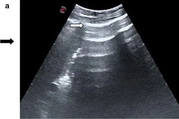
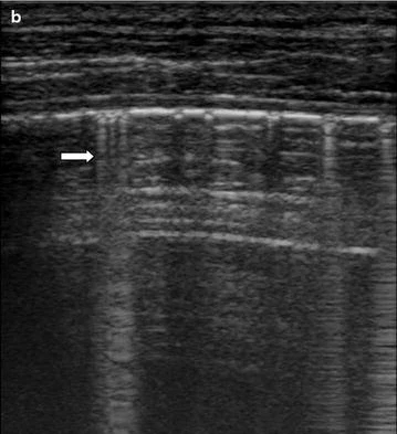
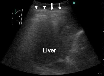
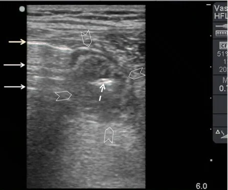
影像來源:Nazerian 2015(Crit Ultrasound J)、Kameda 2016(J Intensive Care)、Abu-Zidan 2018(World J Emerg Surg),裁切單格,CC BY 4.0。
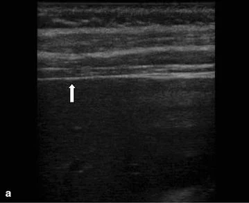
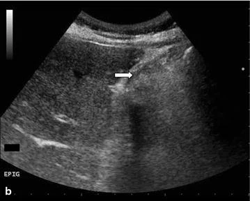
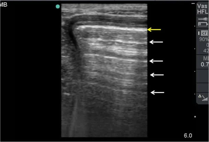
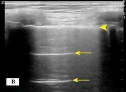
對照:前兩張是沒有游離氣體的正常影像,後兩張是容易誤認的假象。迴響線的樣子和游離氣體很像,差別在氣體的位置(腸腔內、橫膈上方)與會不會隨姿勢移動。
05游離液體
游離液體
- 去哪裡找:右上腹莫氏囊(Morison pouch)、左上腹脾臟周圍、骨盆腔,和創傷 FAST 相同的位置。
- 看回音:完全黑(無回音)可以是腹水、尿液或早期出血;有回音、雜亂的液體要想到血塊或膿(本課程講義)。
- 非創傷病人也會出血:Kameda 2016 的病例沒有外傷記憶,超音波在莫氏囊、脾周與直腸膀胱窩看到液體,CT 證實脾臟破裂。
- 掃描手法見 eFAST 一章。
黃體破裂(本課程講義)
- 囊壁破損或囊腫塌陷。
- 囊腫內沒有都卜勒血流。
- 液體性質:無回音代表漿液或早期出血;複雜、有回音的液體提示血液或血塊。
- 育齡女性有骨盆腔積液,一定先驗孕,排除子宮外孕。
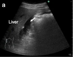
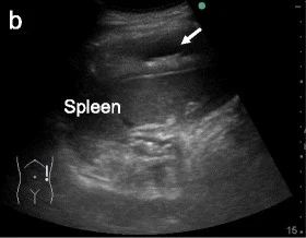
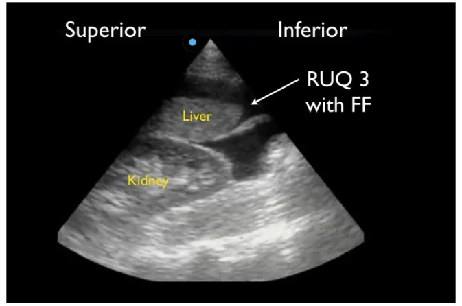
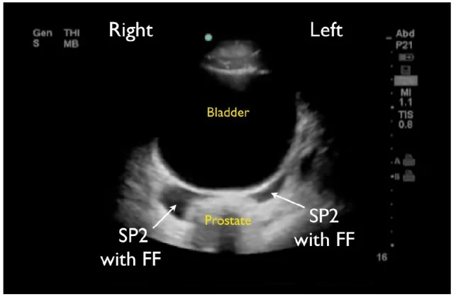
影像來源:Kameda 2016(前兩張)、Lobo 2017(後兩張,創傷病人的 FAST 影像,與 eFAST 章節共用),CC BY 4.0。
以下內容需要免費訂閱
病例影像判讀與練習題開放給訂閱者。留下電子郵件即可立即閱讀全部內容;同一個瀏覽器一年內不必再輸入。換手機或電腦時,輸入同一個信箱即可。
不收費、不寄廣告。若想收到新章節通知,我們會另寄一封確認信,點了才會開始寄。詳見隱私說明。
10參考文獻與延伸閱讀
- Nazerian P, Tozzetti C, Vanni S, et al. Accuracy of abdominal ultrasound for the diagnosis of pneumoperitoneum in patients with acute abdominal pain: a pilot study. Crit Ultrasound J. 2015;7(1):15. doi:10.1186/s13089-015-0032-6
- Laméris W, van Randen A, van Es HW, et al. Imaging strategies for detection of urgent conditions in patients with acute abdominal pain: diagnostic accuracy study. BMJ. 2009;338:b2431. doi:10.1136/bmj.b2431
- Stoker J, van Randen A, Laméris W, Boermeester MA. Imaging patients with acute abdominal pain. Radiology. 2009;253(1):31-46. doi:10.1148/radiol.2531090302
- Smith-Bindman R, Aubin C, Bailitz J, et al. Ultrasonography versus computed tomography for suspected nephrolithiasis. N Engl J Med. 2014;371(12):1100-1110. doi:10.1056/NEJMoa1404446
- Gottlieb M, Peksa GD, Pandurangadu AV, et al. Utilization of ultrasound for the evaluation of small bowel obstruction: a systematic review and meta-analysis. Am J Emerg Med. 2018;36(2):234-242. doi:10.1016/j.ajem.2017.07.085
- Rubano E, Mehta N, Caputo W, Paladino L, Sinert R. Systematic review: emergency department bedside ultrasonography for diagnosing suspected abdominal aortic aneurysm. Acad Emerg Med. 2013;20(2):128-138. doi:10.1111/acem.12080
- Yokoe M, Hata J, Takada T, et al. Tokyo Guidelines 2018: diagnostic criteria and severity grading of acute cholecystitis (with videos). J Hepatobiliary Pancreat Sci. 2018;25(1):41-54. doi:10.1002/jhbp.515
- Wilson SJ, Thavanathan R, Cheng W, et al. Test characteristics of emergency medicine-performed point-of-care ultrasound for the diagnosis of acute cholecystitis: a systematic review and meta-analysis. Ann Emerg Med. 2024;83(3):235-246. doi:10.1016/j.annemergmed.2023.09.005
- Kessler N, Cyteval C, Gallix B, et al. Appendicitis: evaluation of sensitivity, specificity, and predictive values of US, Doppler US, and laboratory findings. Radiology. 2004;230(2):472-478. doi:10.1148/radiol.2302021520
- Stein JC, Wang R, Adler N, et al. Emergency physician ultrasonography for evaluating patients at risk for ectopic pregnancy: a meta-analysis. Ann Emerg Med. 2010;56(6):674-683. doi:10.1016/j.annemergmed.2010.06.563
- Grunau GL, Harris A, Buckley J, Todd NJ. Diagnosis of ovarian torsion: is it time to forget about Doppler? J Obstet Gynaecol Can. 2018;40(7):871-875. doi:10.1016/j.jogc.2017.09.013
- Sato M, Ishida H, Konno K, et al. Chilaiditi syndrome: sonographic findings. Abdom Imaging. 2000;25(4):397-399. doi:10.1007/s002610000020
- Lee SH, Yun SJ. Diagnostic performance of emergency physician-performed point-of-care ultrasonography for acute appendicitis: a meta-analysis. Am J Emerg Med. 2019;37(4):696-705. doi:10.1016/j.ajem.2018.07.025
- Ross M, Brown M, McLaughlin K, et al. Emergency physician-performed ultrasound to diagnose cholelithiasis: a systematic review. Acad Emerg Med. 2011;18(3):227-235. doi:10.1111/j.1553-2712.2011.01012.x
- Gurusamy KS, Giljaca V, Takwoingi Y, et al. Ultrasound versus liver function tests for diagnosis of common bile duct stones. Cochrane Database Syst Rev. 2015;2015(2):CD011548. doi:10.1002/14651858.CD011548
- Wong C, Teitge B, Ross M, Young P, Robertson HL, Lang E. The accuracy and prognostic value of point-of-care ultrasound for nephrolithiasis in the emergency department: a systematic review and meta-analysis. Acad Emerg Med. 2018;25(6):684-698. doi:10.1111/acem.13388
- Wattar B, Rimmer M, Rogozinska E, Macmillian M, Khan KS, Al Wattar BH. Accuracy of imaging modalities for adnexal torsion: a systematic review and meta-analysis. BJOG. 2021;128(1):37-44. doi:10.1111/1471-0528.16371
- Chen SC, Yen ZS, Wang HP, Lin FY, Hsu CY, Chen WJ. Ultrasonography is superior to plain radiography in the diagnosis of pneumoperitoneum. Br J Surg. 2002;89(3):351-354. doi:10.1046/j.0007-1323.2001.02013.x
- Asrani A. Sonographic diagnosis of pneumoperitoneum using the 'enhancement of the peritoneal stripe sign.' A prospective study. Emerg Radiol. 2007;14(1):29-39. doi:10.1007/s10140-007-0583-3
- Gibbons RC, Smith D, Feig R, Mulflur M, Costantino TG. The sonographic protocol for the emergent evaluation of aortic dissections (SPEED protocol): a multicenter, prospective, observational study. Acad Emerg Med. 2024;31(2):112-118. doi:10.1111/acem.14839
- Mitterberger M, Pinggera GM, Colleselli D, et al. Acute pyelonephritis: comparison of diagnosis with computed tomography and contrast-enhanced ultrasonography. BJU Int. 2008;101(3):341-344. doi:10.1111/j.1464-410X.2007.07280.x
圖片來源與授權
- 以下論文的影像在本頁裁切為單格呈現(多格拼圖拆成單格;部分影像遮蓋了機型字樣),圖說為本站中文改寫。
- Nazerian P, Tozzetti C, Vanni S, et al. Accuracy of abdominal ultrasound for the diagnosis of pneumoperitoneum in patients with acute abdominal pain: a pilot study. Crit Ultrasound J 2015;7(1):15, doi:10.1186/s13089-015-0032-6(Fig. 1、2),CC BY 4.0。
- Kameda T, Taniguchi N. Overview of point-of-care abdominal ultrasound in emergency and critical care. J Intensive Care 2016;4:53, doi:10.1186/s40560-016-0175-y(Fig. 1、2、3),CC BY 4.0。
- Abu-Zidan FM, Cevik AA. Diagnostic point-of-care ultrasound (POCUS) for gastrointestinal pathology: state of the art from basics to advanced. World J Emerg Surg 2018;13:47, doi:10.1186/s13017-018-0209-y(Fig. 3、5、6、9),CC BY 4.0。
- Pourmand A, Dimbil U, Drake A, Shokoohi H. The accuracy of point-of-care ultrasound in detecting small bowel obstruction in emergency department. Emerg Med Int 2018;2018:3684081, doi:10.1155/2018/3684081(Figure 1、2),CC BY 4.0。
- Mori T, Shin TS, Ong GYK. High ascending retrocecal appendicitis in a pediatric patient detected by point-of-care ultrasound. Clin Pract Cases Emerg Med 2019;3(2):149-152, doi:10.5811/cpcem.2019.2.41682(Image 2、3),CC BY 4.0。
- McCafferty G, Shorette A, Singh S, Budhram G. Emphysematous pyelonephritis: bedside ultrasound diagnosis in the emergency department. Clin Pract Cases Emerg Med 2017;1(2):92-94, doi:10.5811/cpcem.2016.12.32714(Image 1),CC BY 4.0。
- Varra FN, Fasoulopoulos A, Spathi E, et al. Ultrasonographic whirlpool sign in ovarian torsion with dual mature teratomas: a case report and literature review. Med Int (Lond) 2026;6(4):38, doi:10.3892/mi.2026.322(Figure 3),CC BY 4.0。
- Yang FJ, Kohen B, Sanapala S, Halperin M. A potential pitfall in POCUS of the gallbladder: beware of the duodenum. POCUS J 2022;7(2):188-189, doi:10.24908/pocus.v7i2.15632(Figure 1、2),CC BY 4.0。
- Killu K, Kakol M. Practical Applications of Lung and Diaphragm Ultrasound in the Intensive Care Unit: An Updated Narrative Review. Cureus 2025;17(7):e88584, doi:10.7759/cureus.88584(Fig. 3B),CC BY 4.0。
- Farina R, Vasile T, Foti PV, Pennisi I, Basile A. Wilkie syndrome and pseudo-nutcracker syndrome a rare combination: description of a case. Cureus 2021;13(10):e18612, doi:10.7759/cureus.18612(Figure 3a、3b),CC BY 3.0。
- Ciortescu I, Nemteanu R, Hincu C, Gheorghe L, Plesa A. An underrated diagnosis of superior mesenteric artery syndrome: a case report. Diagnostics (Basel) 2022;12(9):2159, doi:10.3390/diagnostics12092159(Figure 2),CC BY 4.0。
- Genovese EA, Fonio P, Floridi C, et al. Abdominal vascular emergencies: US and CT assessment. Crit Ultrasound J 2013;5(Suppl 1):S10, doi:10.1186/2036-7902-5-S1-S10(Figure 6a),CC BY 2.0。
- Piltcher-da-Silva R, Sasaki VL, Petry JF, et al. Mesenteric venous thrombosis in a pregnant woman at first trimester gestation: a case report. J Surg Case Rep 2022;2022(6):rjac294, doi:10.1093/jscr/rjac294(Figure 4),CC BY 4.0。
- Geyer T, Rübenthaler J, Froelich MF, et al. Contrast-enhanced ultrasound for assessing abdominal conditions in pregnancy. Medicina (Kaunas) 2020;56(12):675, doi:10.3390/medicina56120675(Figure 4a),CC BY 4.0。
- Nahouli L, Chamseddine N, Alameddine O, Aouad G, Ghazeeri G. Spontaneous conception during ovarian stimulation complicated by severe ovarian hyperstimulation syndrome (OHSS) with atypical β-human chorionic gonadotropin (β-hCG) trends: a case report. Cureus 2026;18(4):e107732, doi:10.7759/cureus.107732(Figure 1),CC BY 4.0。
- Azeez M, Porter V. Spontaneous ovarian hyperstimulation syndrome in a naturally conceived singleton pregnancy with familial predisposition: a case report. Cureus 2026;18(9):e115876, doi:10.7759/cureus.115876(Figure 4、5),CC BY 4.0。
- Baqer F, Alsudairy N. Spontaneous ovarian hyperstimulation syndrome in natural conception: a case report. Cureus 2025;17(4):e83110, doi:10.7759/cureus.83110(Figure 1),CC BY 4.0。
- 標示「作者提供」的影像為本站作者自行取得之教學影像,已去除可識別資訊。
- Lobo V, Hunter-Behrend M, Cullnan E, et al. Caudal edge of the liver in the right upper quadrant (RUQ) view is the most sensitive area for free fluid on the FAST exam. West J Emerg Med 2017;18(2):270-280, doi:10.5811/westjem.2016.11.30435(Fig. 4、8),CC BY 4.0。