超音波圖譜 › 第 19 章 · 陰囊與睪丸
陰囊與睪丸超音波
這一章的重點 急性陰囊痛最怕漏掉睪丸扭轉。床邊超音波永遠兩側一起看:比大小、比迴音、比血流,再沿精索往上找扭轉的漩渦。看到血流不代表沒有扭轉;臨床懷疑高時,超音波不能延誤泌尿科會診。
01使用時機:急性陰囊痛
急性陰囊痛是有時間壓力的急症:睪丸扭轉(testicular torsion)要盡快解除,睪丸破裂要早開刀,Fournier 壞疽會危及生命。病史與理學檢查常互相重疊,床邊超音波可以把這幾種狀況分開,但它是加速決策的工具,不是延後會診的理由。
學完這一章,你應該能夠
- 用線性探頭做縱切+橫切,並取得兩側睪丸同框的對照切面。
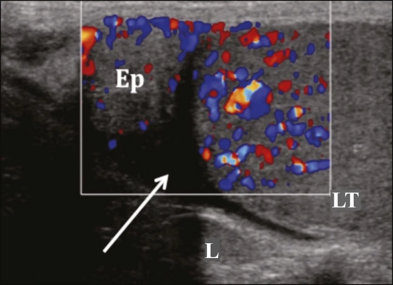
- 認出正常睪丸、睪丸縱隔、副睪頭與白膜。
- 判讀睪丸扭轉:血流不對稱、whirlpool sign、睪丸方向(lie)異常。
- 分辨副睪炎/睪丸炎的充血,與扭轉的缺血。
- 在外傷病人身上找白膜中斷、輪廓變形與血腫;認出 Fournier 壞疽的皮下氣體。
準確度與定位
- 急診醫師做的陰囊超音波:回溯 36 位急性陰囊痛病人,與影像科或手術結果比較,敏感度 95%、特異度 94%(Blaivas 2001)。
- 兒童急診:兒科急診醫師的 POCUS 比影像科超音波結果中位數早 73 分鐘出來;對所有病因的整體一致率 70%,經驗越多一致率越高(Friedman 2019)。
- 扭轉:512 位 ≤15 歲急性陰囊病人,以「睪丸血流減少或 whirlpool sign」為陽性,敏感度 96%、特異度 95%;作者仍強調有不可忽視的偽陰性(Nakamura 2026)。
作者的課程講義把這堂課放在「女性與男性急症」中:男性部分依序談精索 whirlpool sign、精索冗長、睪丸橫躺(horizontal lie)、副睪炎、睪丸外傷與正常睪丸的掃法。本章依講義骨架整理,並補上原始文獻。
02解剖與正常影像
陰囊的層次
- 由外到內:皮膚 → 肉膜(tunica dartos,薄層肌肉)→ 筋膜層;肉膜與筋膜在中線融合成中隔,把左右陰囊隔開。
- 鞘膜(tunica vaginalis)分臟層與壁層,兩層之間是潛在空間——陰囊水腫、血腫、膿液就積在這裡。
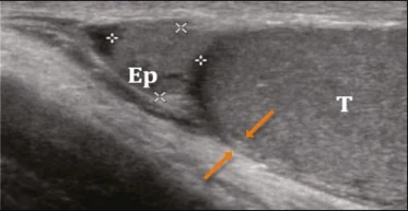
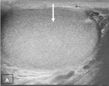
- 睪丸外包一層白膜(tunica albuginea),超音波上是圍著睪丸的高迴音細線;外傷時要找它有沒有斷。
睪丸與精索
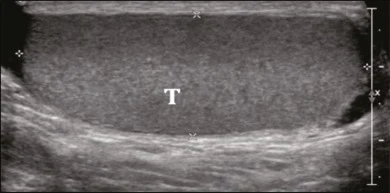
- 白膜向內延伸成睪丸縱隔(mediastinum testis),超音波上是睪丸中央的高迴音線條,並分出小葉間隔,把睪丸分成許多小葉。
- 正常睪丸迴音均勻、細顆粒狀;副睪頭位在睪丸上極,迴音與睪丸相近(Fonseca 2018)。
- 精索含輸精管、睪丸動脈、輸精管動脈、蔓狀靜脈叢、淋巴與神經,外覆精索外筋膜、提睪肌與精索內筋膜。扭轉就是這一束扭在一起。
03掃描技巧:永遠兩側一起看
步驟
- 病人平躺,大腿間或陰囊下方墊毛巾托住陰囊,陰莖往上放在下腹並蓋好。
- 高頻線性探頭,多擠凝膠。先掃沒痛的那側,調好灰階與都卜勒當作基準。
- 每側都做縱切與橫切,從上極掃到下極、內側掃到外側,再看副睪頭、體、尾。
- 探頭橫放在中線,取得兩側同框的對照切面,比較大小、迴音與血流(Mariz 2025)。
- 沿著精索往上掃到腹股溝外環,找扭轉的漩渦或冗長的精索(Kalfa 2007;Bandarkar 2018)。
都卜勒設定
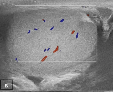
- 睪丸內血流慢:把速度標尺(scale/PRF)調低、增益調到剛好不出雜訊,再比較兩側。
- 兩側一定要用同一組設定,否則「不對稱」可能只是設定不同。
- 能量都卜勒對慢速血流較敏感,可以和彩色都卜勒互補;必要時用頻譜都卜勒確認是動脈波形。
- 記錄:兩側血流是否對稱、睪丸方向、精索型態、有無積液、陰囊壁厚度與氣體。
04睪丸扭轉:血流、漩渦與方向
主要徵象
- 血流不對稱:患側睪丸內血流減少或消失,對側正常。這是最常用的徵象(Mariz 2025)。
- Whirlpool sign:精索扭在一起,橫切像漩渦或蝸牛殼。統合分析合併敏感度 0.73、特異度 0.99;排除新生兒後敏感度升到 0.92(McDowall 2018)。
- 睪丸方向(lie)異常:正常睪丸縱隔朝後外側;扭轉時睪丸可能橫躺或斜躺(Bandarkar 2018)。
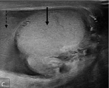
- 其他:睪丸腫大變圓、迴音變得不均勻、精索冗長堆在睪丸上方、反應性積液(Bandarkar 2018)。
都卜勒的侷限
- 歐洲 11 家醫院 208 位手術證實的兒童精索扭轉中,患側睪丸內血流消失只占 76%;直接追精索找扭轉則在 96% 看到(Kalfa 2007)。
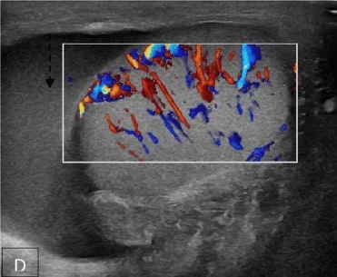
- 部分扭轉、間歇性扭轉或剛自行轉回時,睪丸內仍可能有血流(Bandarkar 2018)。
- 反過來說,彩色都卜勒在 298 位疑似扭轉、全數接受手術的男童的研究中敏感度 96.8%、特異度 97.9%(Waldert 2010)——好的檢查很準,但不是零漏診。
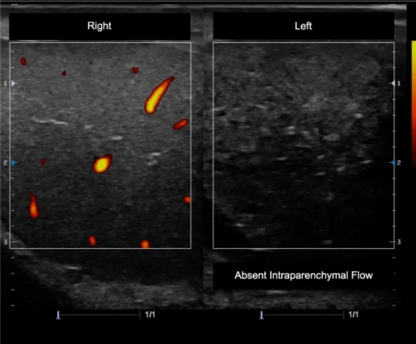
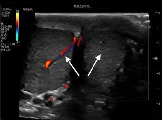
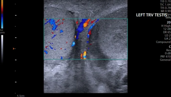
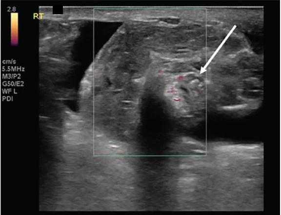
不典型的扭轉:有血流也可能是扭轉
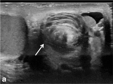
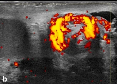
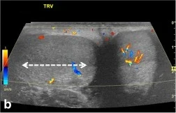
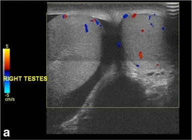
超音波不能延誤會診 病史與理學檢查高度懷疑扭轉時,先通知泌尿科,超音波與會診同時進行。超音波正常或有血流都不能單獨排除扭轉。Nakamura 2026 的結論是 TWIST 分數應用於風險分層、不宜當排除工具,POCUS 也有偽陰性。
床邊徒手復位(manual detorsion)有個案報告在超音波監測下進行,但復位後仍需泌尿科評估與手術固定,本章不展開。
05副睪炎與睪丸炎
影像表現
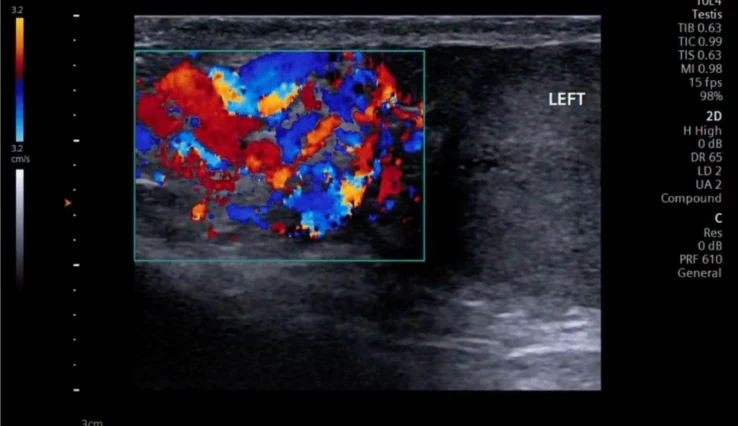
- 副睪腫大、迴音不均、血流增加(Mariz 2025;Kummari 2023)。
- 睪丸受侵犯時(副睪睪丸炎),睪丸也腫大、迴音不均、瀰漫性充血。
- 常伴隨反應性陰囊水腫與陰囊壁增厚;積液內有回音時要想到膿(pyocele)。
和扭轉怎麼分
- 關鍵是血流方向相反:發炎是充血,扭轉是缺血。一定要和對側比。
- 副睪腫大但沒有充血時,要想到扭轉造成的副睪精索團塊,不要直接當副睪炎(Bandarkar 2018)。
- 睪丸附件扭轉:睪丸上極旁一顆腫大、無血流的小結節,旁邊可有反應性充血,睪丸本身迴音正常(Fonseca 2018)。
以下內容需要免費訂閱
病例影像判讀與練習題開放給訂閱者。留下電子郵件即可立即閱讀全部內容;同一個瀏覽器一年內不必再輸入。換手機或電腦時,輸入同一個信箱即可。
不收費、不寄廣告。若想收到新章節通知,我們會另寄一封確認信,點了才會開始寄。詳見隱私說明。
11參考文獻與延伸閱讀
- Blaivas M, Sierzenski P, Lambert M. Emergency evaluation of patients presenting with acute scrotum using bedside ultrasonography. Acad Emerg Med. 2001;8(1):90-93. doi:10.1111/j.1553-2712.2001.tb00563.x
- Friedman N, Pancer Z, Savic R, et al. Accuracy of point-of-care ultrasound by pediatric emergency physicians for testicular torsion. J Pediatr Urol. 2019;15(6):608.e1-608.e6. doi:10.1016/j.jpurol.2019.07.003
- Nakamura T, Kinoshita M, Ihara T, et al. Evaluating the TWIST score and point-of-care ultrasound for paediatric testicular torsion. Emerg Med J. 2026;43(6):334-340. doi:10.1136/emermed-2025-215067
- Kalfa N, Veyrac C, Lopez M, et al. Multicenter assessment of ultrasound of the spermatic cord in children with acute scrotum. J Urol. 2007;177(1):297-301. doi:10.1016/j.juro.2006.08.128
- Waldert M, Klatte T, Schmidbauer J, et al. Color Doppler sonography reliably identifies testicular torsion in boys. Urology. 2010;75(5):1170-1174. doi:10.1016/j.urology.2009.07.1298
- McDowall J, Adam A, Gerber L, et al. The ultrasonographic "whirlpool sign" in testicular torsion: valuable tool or waste of valuable time? A systematic review and meta-analysis. Emerg Radiol. 2018;25(3):281-292. doi:10.1007/s10140-018-1579-x
- Bandarkar AN, Blask AR. Testicular torsion with preserved flow: key sonographic features and value-added approach to diagnosis. Pediatr Radiol. 2018;48(5):735-744. doi:10.1007/s00247-018-4093-0
- Buckley JC, McAninch JW. Use of ultrasonography for the diagnosis of testicular injuries in blunt scrotal trauma. J Urol. 2006;175(1):175-178. doi:10.1016/S0022-5347(05)00048-0
- Guichard G, El Ammari J, Del Coro C, et al. Accuracy of ultrasonography in diagnosis of testicular rupture after blunt scrotal trauma. Urology. 2008;71(1):52-56. doi:10.1016/j.urology.2007.09.014
- Morrison D, Blaivas M, Lyon M. Emergency diagnosis of Fournier's gangrene with bedside ultrasound. Am J Emerg Med. 2005;23(4):544-547. doi:10.1016/j.ajem.2004.12.010
- Mariz J, Martinez J, Arroja S, Blaivas M. Testicular ultrasound: an emergency medicine perspective. Intern Emerg Med. 2025;20(4):1153-1163. doi:10.1007/s11739-025-03864-z
- Fonseca EKUN, Peixoto MR, Cavalcante Júnior FA, et al. Ultrasound evaluation of inguinoscrotal pain: an imaging-based review for the ultrasonographer. Radiol Bras. 2018;51(3):193-199. doi:10.1590/0100-3984.2016.0175
- Kummari S, Das S, Mahajan S. Role of high-resolution ultrasonography with colour and duplex Doppler in the evaluation of acute scrotal diseases. Cureus. 2023;15(11):e49231. doi:10.7759/cureus.49231
- Papathanassiou ZG, Michalopoulou K, Alexopoulos V, Panagidis A. Testicular rupture: clinical, sonographic, and surgical correlation in an adolescent patient. Cureus. 2025;17(2):e78688. doi:10.7759/cureus.78688
- Ramm L, Guidry K, Cirilli A, Kurkowski E, Yu C. Critical point-of-care ultrasound diagnosis of Fournier's gangrene: a case report. Clin Pract Cases Emerg Med. 2022;6(1):57-60. doi:10.5811/cpcem.2021.11.54849
圖片來源與授權
- 以下論文的影像在本頁裁切為單格呈現(多格拼圖拆成單格;部分影像裁去或以黑塊遮去機器上的廠牌/機型字樣),圖說為本站中文改寫。
- Fonseca EKUN, Peixoto MR, Cavalcante Júnior FA, et al. Ultrasound evaluation of inguinoscrotal pain: an imaging-based review for the ultrasonographer. Radiol Bras 2018;51(3):193-199, doi:10.1590/0100-3984.2016.0175(Fig. 2、3、7、11),CC BY 4.0。
- Kummari S, Das S, Mahajan S. Role of High-Resolution Ultrasonography With Colour and Duplex Doppler in the Evaluation of Acute Scrotal Diseases. Cureus 2023;15(11):e49231, doi:10.7759/cureus.49231(Fig. 5、7),CC BY 4.0。
- Mariz J, Martinez J, Arroja S, Blaivas M. Testicular ultrasound: an emergency medicine perspective. Intern Emerg Med 2025;20(4):1153-1163, doi:10.1007/s11739-025-03864-z(Fig. 1、2、4、5、6),CC BY 4.0。
- Small E, Ashenburg N, Schertzer K. 19-Year-Old with Sudden Onset Left Testicular Pain. Clin Pract Cases Emerg Med 2022;6(4):327-329, doi:10.5811/cpcem.2022.7.56747(Image 1),CC BY 4.0。
- Correa C, Onishi S, Abrams E. Testicular Torsion in Monorchism Diagnosed with Point-of-care Ultrasound: A Case Report. Clin Pract Cases Emerg Med 2021;5(1):82-84, doi:10.5811/cpcem.2020.12.48944(Image 1、3),CC BY 4.0。
- Bandarkar AN, Blask AR. Testicular torsion with preserved flow: key sonographic features and value-added approach to diagnosis. Pediatr Radiol 2018;48(5):735-744, doi:10.1007/s00247-018-4093-0(Fig. 2、5、10),CC BY 4.0。
- Holliday TL, Robinson KS, Dorinzi N, et al. Testicular Rupture: A Tough Nut to Crack. Clin Pract Cases Emerg Med 2017;1(3):221-224, doi:10.5811/cpcem.2017.3.33348(Image 2),CC BY 4.0。
- Papathanassiou ZG, Michalopoulou K, Alexopoulos V, Panagidis A. Testicular Rupture: Clinical, Sonographic, and Surgical Correlation in an Adolescent Patient. Cureus 2025;17(2):e78688, doi:10.7759/cureus.78688(Fig. 1),CC BY 4.0。
- Ramm L, Guidry K, Cirilli A, Kurkowski E, Yu C. Critical Point-of-care Ultrasound Diagnosis of Fournier's Gangrene: A Case Report. Clin Pract Cases Emerg Med 2022;6(1):57-60, doi:10.5811/cpcem.2021.11.54849(Image),CC BY 4.0。