急診經食道心臟超音波
01為什麼要用經食道超音波
心跳停止時,經胸心臟超音波(TTE)要在暫停壓胸的幾秒內找窗口,病人插管、肥胖、肺氣腫或接上機械壓胸機時常常看不到。經食道心臟超音波(TEE)探頭在食道裡,緊貼心臟後方,不受胸壁與壓胸干擾,可以在壓胸中持續看,也可以停在同一個切面反覆比較(Teran 2020)。
心跳停止的四件事
- 有沒有心臟活動:真正停滯、假性無脈搏電氣活動(pseudo-PEA)或細微心室顫動(fine VF)。
- 可逆病因:心包填塞、右心擴大、主動脈剝離、低血容。
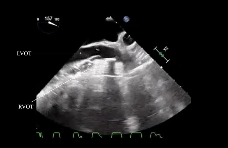
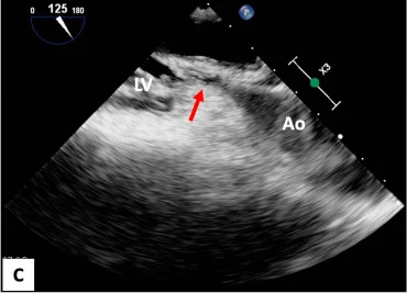
- 壓胸品質:看壓胸最大受壓區(area of maximal compression,AMC)在左心室還是在主動脈根部/左心室出口(LVOT)。
- 縮短暫停:脈搏檢查時探頭已在位,看一眼就繼續壓。
休克與重症
- TTE 窗口不足時,用來找休克原因與連續監測左右心功能。
- 插管病人的輸液反應評估(上腔靜脈隨呼吸的變化)。
- 不明原因低血氧、懷疑主動脈剝離或肺栓塞、導引葉克膜(ECMO)導絲與管路位置。
禁忌
- 食道損傷或狹窄、近期食道或胃手術;懷疑食道損傷的頸部穿刺傷不要放。
- 沒有確定的呼吸道:急診 TEE 原則上在氣管插管後才放。
- 相對禁忌:食道靜脈瘤、活動性上消化道出血,要權衡利弊。
訓練原則:先有 TTE 基礎,再上 TEE 課程與模擬器練習。ACEP 指引建議至少完成 10 次在病人或模擬器上的 TEE 檢查(Fair 2018),能力評核方式由各院自訂。
02探頭、術語與置入
四個深度、五種操作
深度以門齒起算(教科書常用參考值):上食道(UE)約 20–30 公分、中食道(ME)約 30–40 公分、經胃(TG)約 40–45 公分,再往前是深經胃(deep TG),實際依體型調整。
操作只有五種:前送/後退、左轉/右轉(轉整支探頭)、前屈/後屈(大輪)、左右側屈(小輪)、多平面角度(omniplane,按鈕 0–180°)。急診 TEE 主要靠前送後退、轉向、前屈與角度。
置入步驟(已插管)
- 確認氣管內管位置與固定,放咬口器;探頭塗凝膠,套保護套。
- 兩個轉輪都放開(unlocked)、回到中立位,探頭面朝前(朝舌頭)。
- 抬下顎(jaw thrust)或用喉頭鏡把舌根撐開,沿中線輕送。
- 有阻力不要硬推:退一點、調整頭頸角度,或在喉頭鏡直視下再試。
- 通常不必為了放探頭停止壓胸。
| 操作 | 原文(Hahn 2013) | 怎麼做 | 急診常見用途 |
|---|---|---|---|
| ① 前送/後退 | Advance / Withdraw | 整支探頭沿食道推進或拉出 | 換深度:UE → ME → TG |
| ② 左轉/右轉 | Turn to the right (clockwise) / to the left (counterclockwise) | 握住探頭本體,整支繞長軸轉 | ME4C 右轉看右心與三尖瓣、左轉看左心;Bicaval 需右轉 |
| ③ 前屈/後屈 | Anteflex / Retroflex | 轉大輪,尖端往前(心臟側)或往後彎 | 經胃切面靠前屈讓探頭貼住胃壁 |
| ④ 左右側屈 | Flex to the right / left | 轉小輪,尖端往左或往右彎 | 微調;急診較少用 |
| ⑤ 多平面角度 | Rotate forward / back (0–180°) | 按探頭握把上的按鈕,影像平面在探頭內轉動 | ME4C 0–10° → Bicaval 90–110° → ME LAX 120–140° |
術語依 Hahn RT, et al. ASE/SCA 2013 TEE 指引(見參考文獻)。
03四個核心切面
ACEP 指引建議急診心跳停止用四個切面:中食道四腔(ME4C)、中食道長軸(ME LAX)、經胃短軸(TG SAX)與中食道雙腔靜脈(ME bicaval)(Fair 2018)。切面命名依 ASE/SCA 指引(Hahn 2013)。
| 切面 | 怎麼到 | 看什麼 |
|---|---|---|
| ME4C | 中食道 0–10°;心尖看不到時稍後屈或後退 | 四個腔室大小比例、左右心收縮、心包積液、心律(顫動或停滯) |
| ME LAX | 從 ME4C 角度轉到 120–140° | 左心室、LVOT、主動脈瓣與主動脈根部;壓胸時找 AMC |
| TG SAX | 回 0°、前送入胃,前屈貼胃壁 | 乳頭肌層左心室短軸:整體收縮、局部室壁運動、右心壓迫(D 字形)、容積 |
| ME bicaval | ME4C 角度轉到 90–110°,整支探頭右轉 | 上下腔靜脈與右心房;輸液反應、ECMO 導絲與管路位置、心房中隔 |
建議順序:ME4C → ME LAX → ME4C → TG SAX → ME4C → bicaval,每次都回到 ME4C 這個「家」,迷路時最容易重新定位。
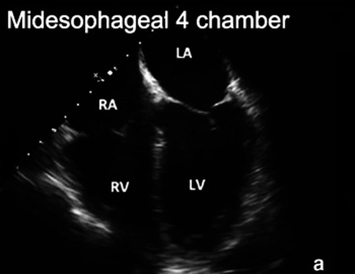
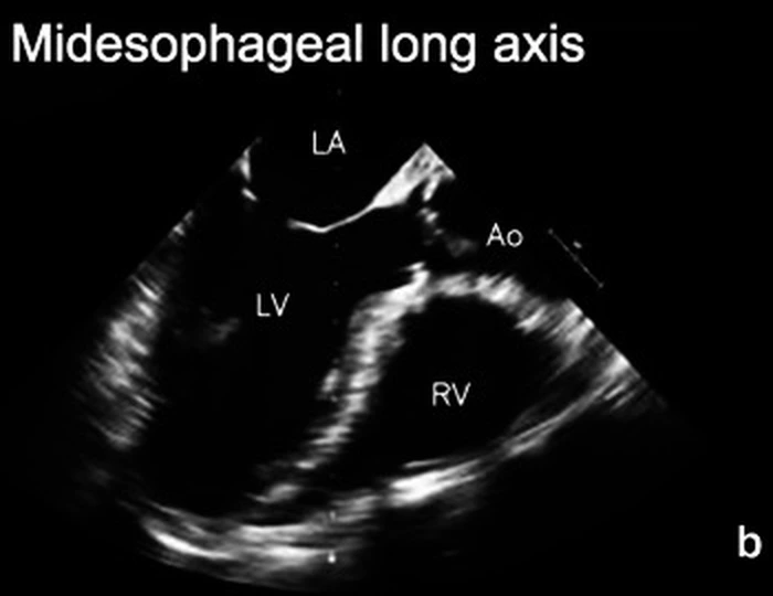
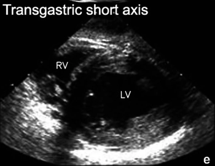
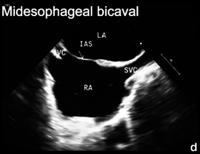
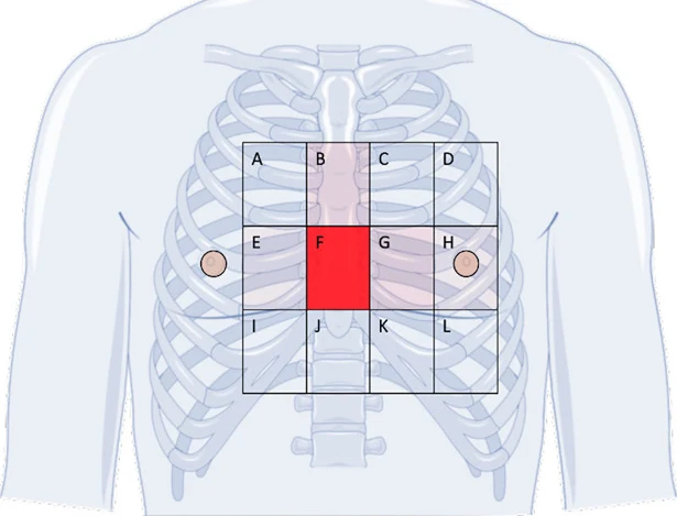
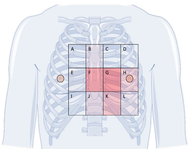
四張圖出自同一個急診心跳停止 TEE 簡化流程的教學研究(Nazerian 2020);圖說的「用途」依原文改寫,指這個切面能回答的問題,不代表圖中有該病灶。
04CPR 中的 TEE
流程:插管後就放
- 插管前:暫停壓胸時用 TTE(劍突下)快速看心包填塞。
- 插管後:放 TEE,壓胸中先到 ME4C 看心臟與心律,再到 ME LAX 找 AMC。
- 脈搏檢查前就把切面擺好,暫停時只看、不找;錄短片,回頭再判讀。
- 找可逆病因(下一節),並持續監測壓胸效果與恢復自發循環(ROSC)。
壓在哪裡:左心室還是 LVOT?
按照標準位置壓胸時,最大受壓區常落在主動脈根部或 LVOT,而不是左心室:34 位病人中 AMC 在主動脈 59%、LVOT 41%,且全部都有出口狹窄(Hwang 2009);急診到院前心跳停止的 TEE 研究中,53% 的 AMC 在主動脈根部或 LVOT(Teran 2019)。
觀察性研究中,主動脈瓣未被壓到的病人持續 ROSC 較多(53.8% vs 24.3%),存活出院則無差異(Chu 2024)。單一中心群集隨機試驗(EXECT-CPR,132 人)用 TEE 調整按壓位置,持續 ROSC 44% vs 39%,未達統計差異,作者說明樣本數不足(Chu 2026)。
目前證據顯示 TEE 引導可改善壓胸當下的血流指標,但還沒有證明改善存活;它是一個看得見的回饋工具,不是保證。
以下內容需要免費訂閱
病例影像判讀與練習題開放給訂閱者。留下電子郵件即可立即閱讀全部內容;同一個瀏覽器一年內不必再輸入。換手機或電腦時,輸入同一個信箱即可。
不收費、不寄廣告。若想收到新章節通知,我們會另寄一封確認信,點了才會開始寄。詳見隱私說明。
08參考文獻與延伸閱讀
- Fair J, Mallin M, Mallemat H, Zimmerman J, Arntfield R, Kessler R, Bailitz J, Blaivas M. Transesophageal echocardiography: guidelines for point-of-care applications in cardiac arrest resuscitation. Ann Emerg Med. 2018;71(2):201-207. doi:10.1016/j.annemergmed.2017.09.003
- Hahn RT, Abraham T, Adams MS, Bruce CJ, et al. Guidelines for performing a comprehensive transesophageal echocardiographic examination: recommendations from the American Society of Echocardiography and the Society of Cardiovascular Anesthesiologists. J Am Soc Echocardiogr. 2013;26(9):921-964. doi:10.1016/j.echo.2013.07.009
- Mayo PH, Narasimhan M, Koenig S. Critical care transesophageal echocardiography. Chest. 2015;148(5):1323-1332. doi:10.1378/chest.15-0260
- Teran F, Prats MI, Nelson BP, Kessler R, et al. Focused transesophageal echocardiography during cardiac arrest resuscitation: JACC review topic of the week. J Am Coll Cardiol. 2020;76(6):745-754. doi:10.1016/j.jacc.2020.05.074
- Arntfield R, Pace J, Hewak M, Thompson D. Focused transesophageal echocardiography by emergency physicians is feasible and clinically influential: observational results from a novel ultrasound program. J Emerg Med. 2016;50(2):286-294. doi:10.1016/j.jemermed.2015.09.018
- Teran F, Dean AJ, Centeno C, Panebianco NL, Zeidan AJ, Chan W, Abella BS. Evaluation of out-of-hospital cardiac arrest using transesophageal echocardiography in the emergency department. Resuscitation. 2019;137:140-147. doi:10.1016/j.resuscitation.2019.02.013
- Fair J 3rd, Mallin MP, Adler A, Ockerse P, Steenblik J, Tonna J, Youngquist ST. Transesophageal echocardiography during cardiopulmonary resuscitation is associated with shorter compression pauses compared with transthoracic echocardiography. Ann Emerg Med. 2019;73(6):610-616. doi:10.1016/j.annemergmed.2019.01.018
- Hwang SO, Zhao PG, Choi HJ, Park KH, Cha KC, Park SM, Kim SC, Kim H, Lee KH. Compression of the left ventricular outflow tract during cardiopulmonary resuscitation. Acad Emerg Med. 2009;16(10):928-933. doi:10.1111/j.1553-2712.2009.00497.x
- Chu SE, Huang CY, Cheng CY, Chan CH, et al. Cardiopulmonary resuscitation without aortic valve compression increases the chances of return of spontaneous circulation in out-of-hospital cardiac arrest: a prospective observational cohort study. Crit Care Med. 2024;52(9):1367-1379. doi:10.1097/CCM.0000000000006336
- Chu SE, Cheng CY, Chang CJ, Ho H, et al. Transesophageal echocardiography during CPR in patients with out-of-hospital cardiac arrest: the EXECT-CPR randomized clinical trial. JAMA Intern Med. 2026;186(5):557-566. doi:10.1001/jamainternmed.2026.0102
- Chen CC, Chen TY, Lin LC, Wu CY, Wu MC, Hsu SH, Tay J, Lien WC. Hemodynamic effects of TEE-guided adjustment of the area of the maximal compressions during cardiopulmonary resuscitation. Resuscitation. 2026:111097. doi:10.1016/j.resuscitation.2026.111097
- Chen CC. Bidirectional within-patient changes in cerebral oxygenation with 3-dimensional transesophageal echocardiography-guided repositioning of the compression site during mechanical cardiopulmonary resuscitation. J Cardiothorac Vasc Anesth. 2026 (online ahead of print). doi:10.1053/j.jvca.2026.08.129
圖片來源與授權
- 以下論文的多格拼圖在本頁拆成單格裁切呈現,圖說為本站中文改寫。授權依 PMC Open Data 的 license_code 核對。
- Amador Y. Point of care transesophageal echocardiogram-guided CPR: area of maximal compression. POCUS J 2025;10(1):61-63, doi:10.24908/pocusj.v10i01.18111(Fig. 1),CC BY 4.0。
- Bernard S, et al. Physiology-guided resuscitation: monitoring and augmenting perfusion during cardiopulmonary arrest. J Clin Med 2024;13(12):3527, doi:10.3390/jcm13123527(Fig. 2A–C),CC BY 4.0。
- Marshall RA, et al. Transesophageal echocardiography during cardiopulmonary resuscitation is associated with alternate areas of compression. PLoS One 2026;21(1):e0339974, doi:10.1371/journal.pone.0339974(Fig. 1),CC BY 4.0。
- Krammel M, et al. Feasibility of resuscitative transesophageal echocardiography at out-of-hospital emergency scenes of cardiac arrest. Sci Rep 2023;13:20085, doi:10.1038/s41598-023-46684-x(Fig. 2、3),CC BY 4.0。
- Chen H, Zhu T. Cardiac arrest due to tamponade during secondary-stage endovascular stent implantation in a patient with DeBakey type I dissection. Front Med (Lausanne) 2026;13:1815531, doi:10.3389/fmed.2026.1815531(Fig. 2A、B),CC BY 4.0。
- Burton E, et al. Left ventricular outflow tract obstruction from proximal intimo-intimal intussusception: a fatal complication of acute type A aortic dissection. Cureus 2026;18(9):e116796, doi:10.7759/cureus.116796(Fig. 1、2),CC BY 4.0。
- Andrade H, et al. Hemodialysis catheter-associated right atrial thrombus diagnosed via point of care transesophageal echocardiogram. POCUS J 2024;9(1):25-28, doi:10.24908/pocus.v9i1.16895(Fig. 4),CC BY 4.0。
- Tu YC, et al. Aortic dissection mimicry under extracorporeal membrane oxygenation (ECMO) after cardiac arrest. Reports 2026;9(3):262, doi:10.3390/reports9030262(Fig. 2),CC BY 4.0。
- Smart KJ, et al. Extracardiac tamponade and hemoperitoneum after cardiopulmonary resuscitation: multimodal diagnosis using point-of-care ultrasonography and transesophageal echocardiography. Cureus 2026;18(8):e114219, doi:10.7759/cureus.114219(Fig. 4A、B),CC BY 4.0。
- Nazerian P, De Stefano G, Albano G, et al. Transesophageal echocardiography (TEE) in cardiac arrest: results of a hands-on training for a simplified TEE protocol. Ultrasound J. 2020;12:41, doi:10.1186/s13089-020-00189-0(Fig. 1a、b、d、e),CC BY 4.0。