超音波圖譜 › 第 27 章 · 超音波導引處置
27.1 超音波導引肋膜積液穿刺與胸管
這一章的重點 肋膜積液穿刺最常見的併發症是氣胸,最危險的是肋間動脈出血。超音波要回答四件事:有沒有夠多的液體、整個呼吸週期裡肺和橫膈會跑到哪、針要走多深、路徑上有沒有血管;做完再看一次 lung sliding。
01使用時機與為什麼要用超音波
共通原則(無菌、in-plane/out-of-plane、靜態定位與即時導引)請看母頁:第 27 章 超音波導引處置。肋膜積液、窗簾徵象、胸椎徵象與氣胸的判讀細節請看肺超音波章。
急診的常見情境
- 診斷性穿刺:新出現、原因不明的肋膜積液,或懷疑感染(肺炎旁積液、膿胸)。
- 治療性穿刺:大量積液造成呼吸困難。
- 胸管/pigtail 引流:氣胸、血胸、複雜性肺炎旁積液或膿胸。
- 不適合當場做:積液太少、找不到安全窗、病人無法維持姿勢;先處理凝血與抗凝問題,或改由專科/影像導引。
超音波降低氣胸 24 篇研究、6,605 次穿刺的統合分析:整體氣胸率 6.0%,其中 34.1% 需放胸管;使用超音波與較低的氣胸風險相關(OR 0.3,95% CI 0.2–0.7)(Gordon 2010)。大型住院資料庫研究中,超音波導引的氣胸風險校正後下降 19%(OR 0.81)(Mercaldi 2013)。
臨床定位常常不準 肺科病房的前瞻研究:醫師依理學檢查選的穿刺點,有 15% 經超音波檢查不準確;醫師找不到穿刺點的情況中,超音波又找出 54% 可穿刺的位置。資深醫師並沒有比訓練中醫師準(Diacon 2003)。
02找積液、判斷複雜度
先確認是肋膜積液
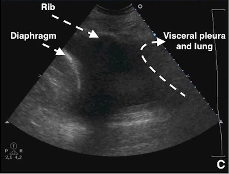
- 低頻凸陣探頭,從後腋線肺底往上掃;先找到橫膈與下方的肝或脾,再看橫膈上方。
- 肋膜積液在橫膈上方呈無回音或低回音區;胸椎影像可一路延伸到橫膈上方(胸椎徵象);肺底的「窗簾」不完整或消失。
- M-mode 上臟層胸膜隨呼吸往胸壁靠近、遠離(正弦波徵象),代表液體可流動。
- 細節與更多圖片見肺超音波:肋膜積液。
量多少?
大小只能粗估。呼吸器病人仰臥、床頭抬高約 15 度,在後腋線量吐氣末壁層到臟層胸膜的最大距離(Sep),V(mL)≈ 20 × Sep(mm),平均預測誤差約 158 mL(Balik 2006)。實務上更重要的是:穿刺點的液體夠不夠厚、離肺與橫膈夠不夠遠。
單純或複雜
- 無回音:漏出液、滲出液都可能,外觀不能取代積液分析。
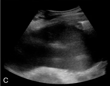
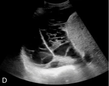
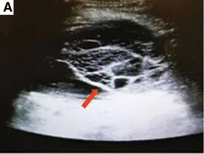
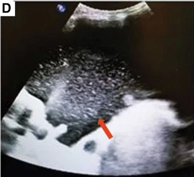
- 有回音、漂浮光點:細胞、蛋白質或碎屑,屬複雜性積液。
- 分隔(septation)、多房(multiloculated):單點穿刺可能抽不乾淨;感染情境要考慮引流的時機與方式。
超音波看得到分隔 SHM 立場聲明建議用超音波偵測分隔等複雜特徵,來決定肋膜引流的時機與方式(Dancel 2018)。
03定位:姿勢、安全三角與呼吸週期
姿勢
- 可以坐:坐姿、身體前傾趴在床邊桌上,穿刺點選後胸或後外側、肋膈角上方液體最厚處。
- 不能坐(重症、插管):仰臥床頭抬高 30–45 度,或患側朝上側臥,在後外側的肋膈角附近穿刺;一項兩院可行性研究在這兩種姿勢下,沿肋膈角以即時超音波導引完成 101 次穿刺與 30 次 pigtail 置放,每次都能即時看到針尖進入肋膜腔(Soldati 2013)。
- 胸管:多選腋中線附近的安全三角(見右欄)。
安全窗要看整個呼吸週期
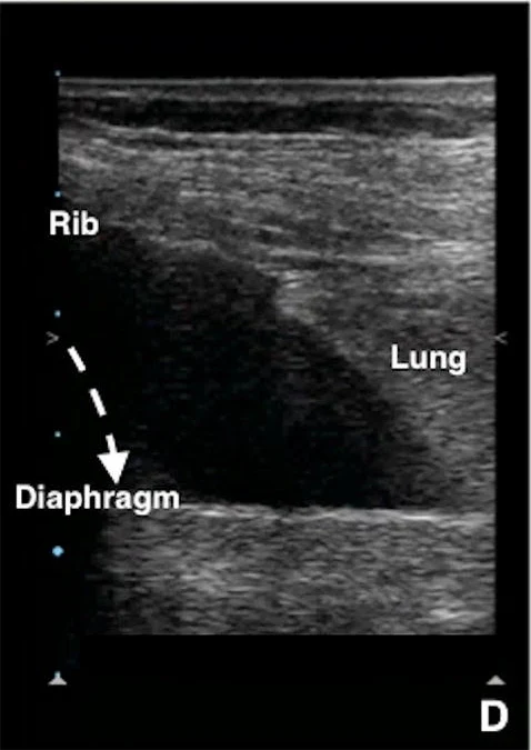
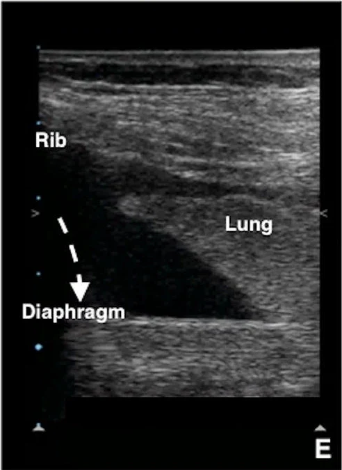
- 吐氣末橫膈最高、吸氣末肺最往下:穿刺點要在兩者之外,整個呼吸週期都看得到液體(Dancel 2018)。
- 探頭放在兩根肋骨之間(橫向),找到壁層與臟層胸膜距離最大的位置,安全距離最大。
- 用超音波量皮膚到壁層胸膜的深度,決定針的長度與最大進針深度(Dancel 2018)。
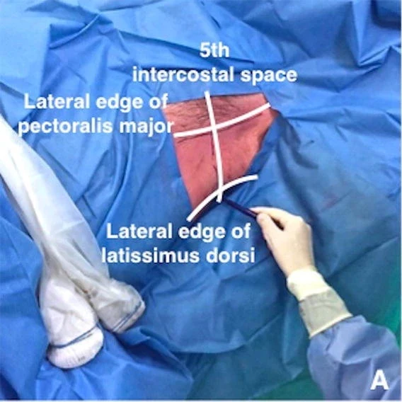
安全三角(safe triangle) 胸管建議放在由胸大肌外緣、闊背肌前緣、乳頭高度的水平線圍成、頂點在腋下的三角區內,以減少傷及下方重要結構的風險(Laws 2003;Havelock 2010)。超音波再確認這個區域內肋間有足夠的液體或確實是氣胸。
標記後不要再動 靜態定位(先畫記號再穿刺)時,標記與穿刺之間不要延遲,病人也不要換姿勢;姿勢一變,液體、肺與橫膈的位置都跟著變(Dancel 2018)。最穩當的是即時導引。
指引怎麼說 英國胸腔學會 2010 年肋膜處置指引建議以胸腔超音波導引肋膜積液的處置(Havelock 2010);2023 年版肋膜疾病指引整合了積液的診斷與處置流程(Roberts 2023)。
04肋間動脈:進針前先開彩色都卜勒
「沿肋骨上緣進針」不一定安全
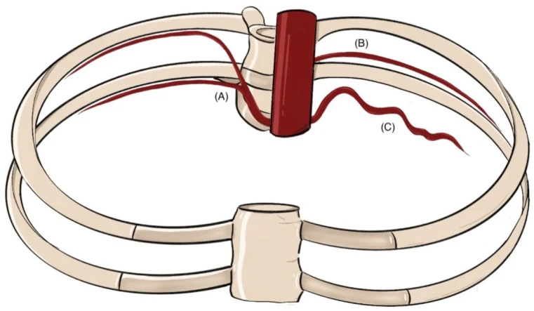
- 傳統教「貼著下一根肋骨上緣進針」,假設肋間動脈藏在上一根肋骨下緣的肋溝內。
- CT 研究(47 位病人、298 條動脈):在脊椎旁 3 cm 處只有 17% 的肋間動脈被上方肋骨遮住,6 cm 處則有 97%;離脊椎 6 cm 內,動脈常暴露在肋間。年紀越大、越上位的肋間,位置越多變(Helm 2013)。
- 超音波研究:越往後(靠脊椎),肋間動脈越靠近肋間正中;但個體差異大,有些人到了腋下仍暴露在肋間(Salamonsen 2012)。
怎麼找
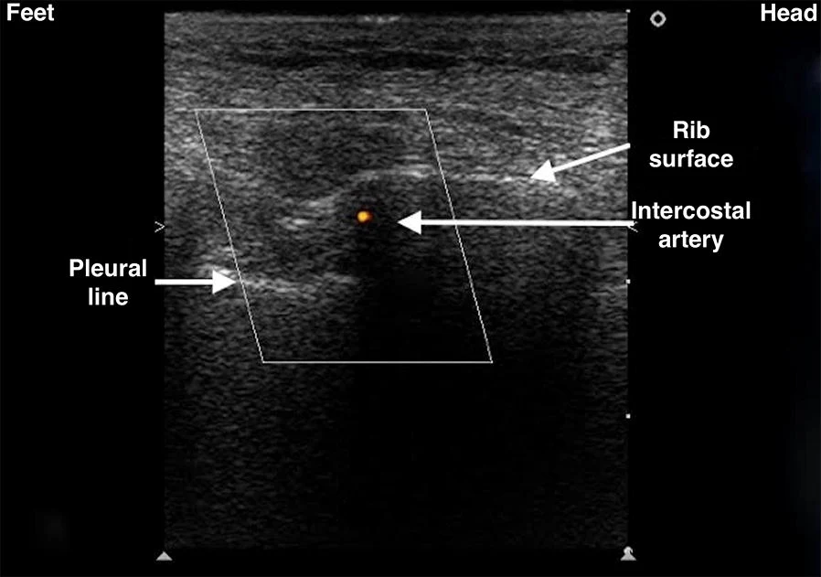
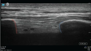
- 高頻線性探頭,沿身體長軸放在預定的肋間,看到上下兩根肋骨與胸膜線。
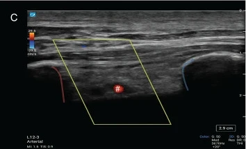
- 彩色都卜勒框放在上方肋骨的下緣,深度調淺到肋骨不要占滿畫面。
- 調低 PRF(脈衝重複頻率)直到看到搏動訊號(Vetrugno 2018 圖說)。
- 動脈若在肋間中間,換一個肋間或往外側移。
篩檢工具,不是保證 與 CT 對照,手提式超音波偵測「暴露在肋間的動脈」敏感度 0.86,但特異度 0.30、陰性預測值 0.27——看不到不代表沒有。原作者建議出血風險較高的病人,穿刺或放胸管前先用超音波篩檢(Salamonsen 2013)。
以下內容需要免費訂閱
病例影像判讀與練習題開放給訂閱者。留下電子郵件即可立即閱讀全部內容;同一個瀏覽器一年內不必再輸入。換手機或電腦時,輸入同一個信箱即可。
不收費、不寄廣告。若想收到新章節通知,我們會另寄一封確認信,點了才會開始寄。詳見隱私說明。
09參考文獻與延伸閱讀
- Gordon CE, Feller-Kopman D, Balk EM, Smetana GW. Pneumothorax following thoracentesis: a systematic review and meta-analysis. Arch Intern Med. 2010;170(4):332-339. doi:10.1001/archinternmed.2009.548
- Mercaldi CJ, Lanes SF. Ultrasound guidance decreases complications and improves the cost of care among patients undergoing thoracentesis and paracentesis. Chest. 2013;143(2):532-538. doi:10.1378/chest.12-0447
- Diacon AH, Brutsche MH, Solèr M. Accuracy of pleural puncture sites: a prospective comparison of clinical examination with ultrasound. Chest. 2003;123(2):436-441. doi:10.1378/chest.123.2.436
- Dancel R, Schnobrich D, Puri N, et al. Recommendations on the use of ultrasound guidance for adult thoracentesis: a position statement of the Society of Hospital Medicine. J Hosp Med. 2018;13(2):126-135. doi:10.12788/jhm.2940
- Havelock T, Teoh R, Laws D, Gleeson F; BTS Pleural Disease Guideline Group. Pleural procedures and thoracic ultrasound: British Thoracic Society Pleural Disease Guideline 2010. Thorax. 2010;65(Suppl 2):ii61-ii76. doi:10.1136/thx.2010.137026
- Roberts ME, Rahman NM, Maskell NA, et al. British Thoracic Society Guideline for pleural disease. Thorax. 2023;78(Suppl 3):s1-s42. doi:10.1136/thorax-2022-219784
- Laws D, Neville E, Duffy J; British Thoracic Society Pleural Diseases Group. BTS guidelines for the insertion of a chest drain. Thorax. 2003;58(Suppl 2):ii53-ii59. doi:10.1136/thorax.58.suppl_2.ii53
- Helm EJ, Rahman NM, Talakoub O, et al. Course and variation of the intercostal artery by CT scan. Chest. 2013;143(3):634-639. doi:10.1378/chest.12-1285
- Salamonsen M, Ellis S, Paul E, et al. Thoracic ultrasound demonstrates variable location of the intercostal artery. Respiration. 2012;83(4):323-329. doi:10.1159/000330920
- Salamonsen M, Dobeli K, McGrath D, et al. Physician-performed ultrasound can accurately screen for a vulnerable intercostal artery prior to chest drainage procedures. Respirology. 2013;18(6):942-947. doi:10.1111/resp.12088
- Balik M, Plasil P, Waldauf P, et al. Ultrasound estimation of volume of pleural fluid in mechanically ventilated patients. Intensive Care Med. 2006;32(2):318. doi:10.1007/s00134-005-0024-2
- Soldati G, Smargiassi A, Inchingolo R, et al. Ultrasound-guided pleural puncture in supine or recumbent lateral position - feasibility study. Multidiscip Respir Med. 2013;8(1):18. doi:10.1186/2049-6958-8-18
- Vetrugno L, Guadagnin GM, Orso D, et al. An easier and safe affair, pleural drainage with ultrasound in critical patient: a technical note. Crit Ultrasound J. 2018;10(1):18. doi:10.1186/s13089-018-0098-z
圖片來源與授權
- 以下論文的多格拼圖在本頁拆成單格裁切呈現,圖說為本站中文改寫;部分影像上的機型或功能商標文字、日期時間與廠牌圖示已以黑色色塊遮蔽。
- Vetrugno L, Guadagnin GM, Orso D, et al. An easier and safe affair, pleural drainage with ultrasound in critical patient: a technical note. Crit Ultrasound J 2018;10(1):18, doi:10.1186/s13089-018-0098-z(Fig. 2、3、4、5、6),CC BY 4.0。
- Soldati G, Smargiassi A, Inchingolo R, et al. Ultrasound-guided pleural puncture in supine or recumbent lateral position - feasibility study. Multidiscip Respir Med 2013;8(1):18, doi:10.1186/2049-6958-8-18(Fig. 3、5),CC BY 2.0。
- Fraser A, Brenner DS, Coghlan M, et al. The Sound of Safety: DIVOT (Doppler Imaging for Vascular Orientation in Thoracic Procedures) Protocol. POCUS J 2025;10(1):83-87, doi:10.24908/pocusj.v10i01.18071(Fig. 4),CC BY 4.0。
- Marchi G, Cinquini S, Tannura F, et al. Intercostal artery screening with color Doppler thoracic ultrasound in pleural procedures: a potential yet underexplored imaging modality for minimizing iatrogenic bleeding risk in interventional pulmonology. J Clin Med 2025;14(17):6326, doi:10.3390/jcm14176326(Fig. 2),CC BY 4.0。
- Casper KP, Sanchirico PJ, Pfeiffer DC. Intercostal artery pseudoaneurysm following thoracentesis: multi-modal imaging and treatment. BMC Med Imaging 2019;19(1):31, doi:10.1186/s12880-019-0333-5(Fig. 3、4),CC BY 4.0。
- Hwang I, Kim J, Hong P, et al. Ultrasound-fluoroscopy versus ultrasound guidance for catheter drainage of loculated pleural effusions: a retrospective study. Diagnostics (Basel) 2026;16(13):2089, doi:10.3390/diagnostics16132089(Fig. 2),CC BY 4.0。
- Wang T, Du G, Fang L, et al. Value of ultrasonography in determining the nature of pleural effusion: analysis of 582 cases. Medicine (Baltimore) 2022;101(33):e30119, doi:10.1097/MD.0000000000030119(Fig. 1),CC BY 4.0。
- Inchingolo R, Ielo S, Barone R, et al. Ultrasound and intrapleural enzymatic therapy for complicated pleural effusion: a case series with a literature review. J Clin Med 2024;13(15):4346, doi:10.3390/jcm13154346(Fig. 11),CC BY 4.0。
- Maly M, Mokotedi MC, Svobodova E, et al. Interpleural location of chest drain on ultrasound excludes pneumothorax and associates with a low degree of chest drain foreshortening on the antero-posterior chest X-ray. Ultrasound J 2022;14(1):45, doi:10.1186/s13089-022-00296-0(Fig. 1),CC BY 4.0。
- Ramírez-Giraldo C, Rey-Chaves CE, Rodriguez Lima DR. Management of pneumothorax with 8.3-French pigtail catheter: description of the ultrasound-guided technique and case series. Ultrasound J 2023;15(1):1, doi:10.1186/s13089-022-00303-4(Fig. 1),CC BY 4.0。