超音波圖譜 › 第 27 章 · 超音波導引處置
27.3 超音波輔助腰椎穿刺
這一章的重點 超音波在腰椎穿刺前告訴你三件事:從哪裡下針(中線與椎間隙)、針要往哪個角度走、大概要多深。做法以「先掃描、畫記號、再穿刺」(mark and poke)為主;畫完記號後病人不能再動。
01使用時機與證據
超音波導引處置的共通原則請見母頁:第 27 章 超音波導引處置。
適應症(本課程講義)
- 懷疑中樞神經感染(腦膜炎)。
- 懷疑蜘蛛膜下腔出血(SAH)。
- 其他神經疾病的檢查、顱內壓的評估與處置。
- 給予治療或診斷用藥物(鞘內給藥)。
併發症(本課程講義)
- 穿刺失敗、創傷性穿刺(血性腦脊髓液)。
- 硬膜穿刺後頭痛、背痛。
- 脊椎硬膜外血腫、硬膜外膿瘍與腦膜炎。
- 深部結構受傷。
多數併發症與選點、角度與進針路徑、進針次數、進針深度有關,而這幾件事正好是超音波能幫忙的地方。
摸的不一定準 100 位做 MRI 的病人中,麻醉醫師用觸診標記腰椎間隙,只有 29% 標對,51% 實際比自以為的高一節;肥胖時更不準(Broadbent 2000)。
統合分析 12 篇隨機試驗(957 人,成人與兒童):超音波輔助組成功率 90.0%,觸診組 81.4%(OR 2.22);創傷性穿刺 10.7% 比 26.5%,進針次數、所需時間與疼痛分數也較少(Gottlieb 2019)。另一篇涵蓋腰椎穿刺與硬膜外導管的統合分析,失敗風險比 0.21(Shaikh 2013)。
效益不是每個病人都一樣:急診 100 位一般成人的隨機試驗中,超音波定位沒有顯著差異(Peterson 2014);而肥胖病人的小型試驗則顯示失敗較少(Nomura 2007;Li 2022)。實務上,摸不到棘突、肥胖、脊椎變形或已經失敗過的病人最值得先掃。
02超音波告訴你什麼:點、角度、深度
- 點:中線(棘突連線)與目標椎間隙的交點,就是進針點。
- 角度:探頭擺出最清楚椎間隙影像時的角度,就是針要走的角度。
- 深度:從皮膚到黃韌帶/後硬膜的距離,預告針大約要進多深。
急診醫師可以很快學會:76 位急診病人中,所有人都看得到骨性與軟組織結構,87.9% 的掃描在 1 分鐘內取得高品質影像(Ferre 2007)。
探頭與擺位
- 瘦的成人或小孩:高頻線性探頭畫面細緻;體型大、結構深:改用低頻凸陣探頭。
- 側躺或坐姿都可以;側躺時肩膀與骨盆都要垂直床面,不要往前或往後翻(本課程講義)。
- 掃描與穿刺必須在同一個姿勢下完成。
肥胖病人 皮膚到黃韌帶的平均距離:BMI 正常者 44 mm,肥胖者(BMI ≥30)64 mm;肥胖者 74% 仍能用超音波辨認相關結構(Stiffler 2007)。
03橫切面:找出中線
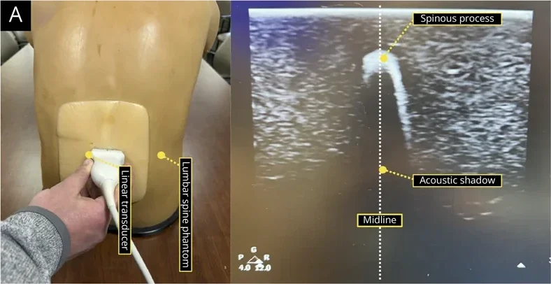
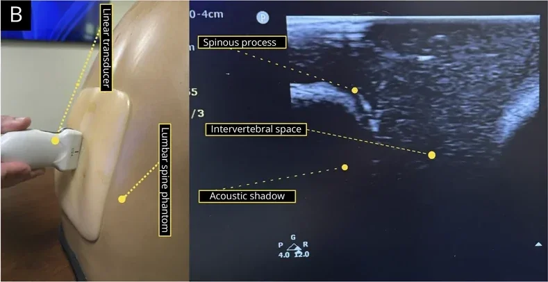
- 探頭橫放在腰椎上,找到棘突:皮下一個亮的尖頂,後方拖著一條黑色聲影。
- 把棘突放在螢幕正中央,在探頭上下緣中點的皮膚各畫一點;上下移動重複幾次,連成中線(棘突連線)。
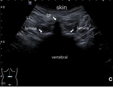
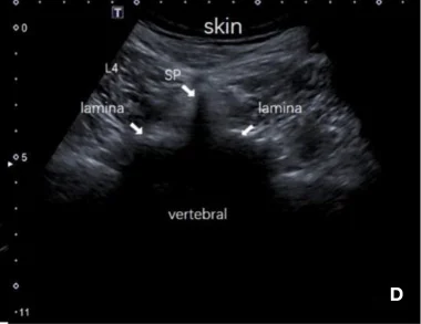
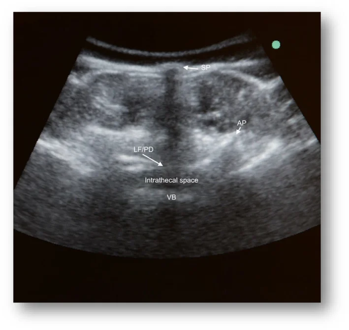
- 探頭稍微往頭側或尾側傾斜,移到兩個棘突之間,可看到橫切椎板間切面(transverse interlaminar view):兩側的關節突、中間的黃韌帶/後硬膜複合體與更深的椎體後緣(前複合體)。
- 看到前、後複合體,代表音束穿過了椎板間的空隙——這就是針可以通過的窗。
04矢狀切面:從薦椎往上數
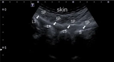
- 探頭轉成縱向、放在中線上,先往尾側找到薦椎:一條連續的亮線。
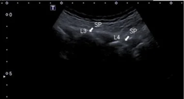
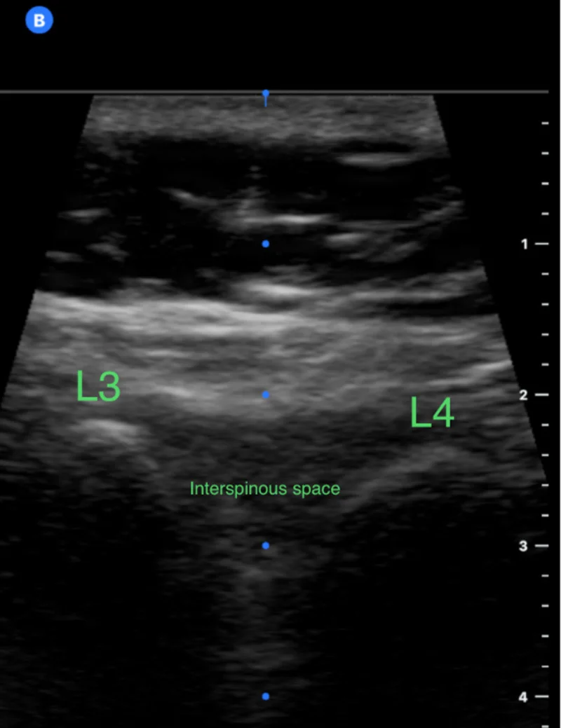
- 往頭側數:L5、L4、L3 棘突呈一個個弧形亮頂,中間的暗區是棘突間隙。
- 把目標間隙(常用 L3–L4 或 L4–L5)放在螢幕中央,在探頭左右兩側中點畫記號。
講義小訣竅:L4、L5、S1 L4 棘突較淺、呈長方形;L5 也呈長方形但較深;S1 則是一條連續的線(本課程講義)。數錯一節,整個定位就跟著錯。
矢狀中線切面的窗口較小,黃韌帶與硬膜不容易分開;要看清楚椎管內的結構,旁正中切面通常較好(Grau 2001)。
以下內容需要免費訂閱
病例影像判讀與練習題開放給訂閱者。留下電子郵件即可立即閱讀全部內容;同一個瀏覽器一年內不必再輸入。換手機或電腦時,輸入同一個信箱即可。
不收費、不寄廣告。若想收到新章節通知,我們會另寄一封確認信,點了才會開始寄。詳見隱私說明。
09參考文獻與延伸閱讀
- Gottlieb M, Holladay D, Peksa GD. Ultrasound-assisted lumbar punctures: a systematic review and meta-analysis. Acad Emerg Med. 2019;26(1):85-96. doi:10.1111/acem.13558
- Shaikh F, Brzezinski J, Alexander S, et al. Ultrasound imaging for lumbar punctures and epidural catheterisations: systematic review and meta-analysis. BMJ. 2013;346:f1720. doi:10.1136/bmj.f1720
- Broadbent CR, Maxwell WB, Ferrie R, et al. Ability of anaesthetists to identify a marked lumbar interspace. Anaesthesia. 2000;55(11):1122-1126. doi:10.1046/j.1365-2044.2000.01547-4.x
- Ferre RM, Sweeney TW. Emergency physicians can easily obtain ultrasound images of anatomical landmarks relevant to lumbar puncture. Am J Emerg Med. 2007;25(3):291-296. doi:10.1016/j.ajem.2006.08.013
- Stiffler KA, Jwayyed S, Wilber ST, et al. The use of ultrasound to identify pertinent landmarks for lumbar puncture. Am J Emerg Med. 2007;25(3):331-334. doi:10.1016/j.ajem.2006.07.010
- Nomura JT, Leech SJ, Shenbagamurthi S, et al. A randomized controlled trial of ultrasound-assisted lumbar puncture. J Ultrasound Med. 2007;26(10):1341-1348. doi:10.7863/jum.2007.26.10.1341
- Peterson MA, Pisupati D, Heyming TW, et al. Ultrasound for routine lumbar puncture. Acad Emerg Med. 2014;21(2):130-136. doi:10.1111/acem.12305
- Li L, Tao W, Cai X. Ultrasound-guided vs. landmark-guided lumbar puncture for obese patients in emergency department. Front Surg. 2022;9:874143. doi:10.3389/fsurg.2022.874143
- Grau T, Leipold RW, Horter J, et al. Paramedian access to the epidural space: the optimum window for ultrasound imaging. J Clin Anesth. 2001;13(3):213-217. doi:10.1016/s0952-8180(01)00245-8
- Wagner J, Dimitrov K, Lauer Z, et al. Increased lumbar puncture success using a paramedian approach: a retrospective cohort study. J Hosp Med. 2026;21(1):42-48. doi:10.1002/jhm.70138
- Conroy PH, Luyet C, McCartney CJ, et al. Real-time ultrasound-guided spinal anaesthesia: a prospective observational study of a new approach. Anesthesiol Res Pract. 2013;2013:525818. doi:10.1155/2013/525818
圖片來源與授權
- 以下論文的多格拼圖在本頁拆成單格裁切呈現,圖說為本站中文改寫。
- Grubman A, Yu A, Bujala N, et al. Ultrasound-assisted lumbar puncture: a narrative review with implications for educators in the clinical neurosciences. Neurol Educ 2026;5(4):e200354, doi:10.1212/NE9.0000000000200354(Figure A、B),CC BY 4.0。
- Li L, Tao W, Cai X. Ultrasound-guided vs. landmark-guided lumbar puncture for obese patients in emergency department. Front Surg 2022;9:874143, doi:10.3389/fsurg.2022.874143(Fig. 3A–D),CC BY 4.0。
- Zanfini BA, Patanella AK, Vassalli F, et al. Ultrasound-assisted and landmark-based nusinersen delivery in spinal muscular atrophy adults: a retrospective analysis. Australas J Ultrasound Med 2024;27(4):210-217, doi:10.1002/ajum.12401(Fig. 1、2),CC BY 4.0。
- Elhassan MG, Mohameden M, Kam M, et al. Feel it, see it, get it: is internal medicine residents' use of ultrasound in lumbar puncture associated with higher success rate? Cureus 2020;12(11):e11545, doi:10.7759/cureus.11545(Fig. 2),CC BY 3.0。
- Gorle Y, Munireddy Papireddy S, Tarigonda S. Evaluation of lumbar sonography as a learning aid for performing subarachnoid block using the paramedian approach by medical junior resident anaesthesiologists: a randomized controlled trial. Cureus 2023;15(5):e38871, doi:10.7759/cureus.38871(Fig. 1),CC BY 3.0。
- Zanfini BA, De Martino S, Frassanito L, et al. "Please mind the gap": successful use of ultrasound-assisted spinal anesthesia for urgent cesarean section in a patient with implanted spinal cord stimulation system for giant chest wall arteriovenous malformation - a case report. BMC Anesthesiol 2020;20(1):122, doi:10.1186/s12871-020-01042-6(Fig. 3),CC BY 4.0。
- Wagner J, Dimitrov K, Lauer Z, et al. Increased lumbar puncture success using a paramedian approach: a retrospective cohort study. J Hosp Med 2026;21(1):42-48, doi:10.1002/jhm.70138(Fig. 1a),CC BY 4.0。
- Backhaus T, von Cranach M, Brich J. Ultrasound-guided lumbar puncture with a needle-guidance system: a prospective and controlled study to evaluate the learnability and feasibility of a newly developed approach. PLoS One 2018;13(4):e0195317, doi:10.1371/journal.pone.0195317(Fig. 1A–D),CC BY 4.0。