超音波圖譜 › 第 27 章 · 超音波導引處置
27.4 超音波導引關節穿刺
這一章的重點 關節穿刺前先用超音波回答兩個問題:真的有關節積液嗎?(還是皮下、滑囊或軟組織的問題)液體在哪裡最多、離皮膚多深、路上有什麼要避開? 懷疑化膿性關節炎時,超音波不能取代穿刺,抽到關節液送檢才是診斷標準。
01為什麼關節穿刺要用超音波
本章共通原則(探頭消毒、in-plane 進針、針尖追蹤)請見母頁 第 27 章 超音波導引處置;關節穿刺前若需要區域止痛,見 超音波導引神經阻斷。
急診的三個用途
- 確認積液:紅腫熱痛的關節,可能是關節積液,也可能是蜂窩性組織炎、滑囊炎或皮下積液。急診回溯研究 54 位病人中,超音波只在 22 位(40.7%)看到關節積液,改變了 35 位(65%)的處置;原本計畫穿刺 39 位,看完超音波後只有 20 位真的穿刺(Adhikari 2010)。
- 懷疑化膿性關節炎:診斷標準是關節穿刺取液檢查(Gottlieb 2019)。超音波負責找到液體、導引穿刺,不能用影像本身判斷有沒有感染。
- 深部或難摸的關節:髖關節、肥胖病人的膝關節、小關節,觸診定位困難,最能從超音波受益。
證據怎麼說
| 研究 | 發現 |
|---|---|
| 風濕科前導研究(Balint 2002) | 傳統方式 32 個關節抽到 10 個(32%);超音波組 31 個關節中 97% 成功,小關節差異最明顯 |
| 急診膝關節 RCT,66 人(Wiler 2010) | 成功率無差異(37/39 vs 25/27),但超音波組較不痛、操作者覺得較容易 |
| 膝關節 RCT,64 膝(Sibbitt 2012) | 超音波導引疼痛較低(VAS 3.0 vs 5.8 cm),抽出液量較多(34 vs 12 mL) |
| 膝骨關節炎統合分析,21 篇(Deng 2025) | 超音波導引的準確度較高(RR 1.26,95% CI 1.09–1.46) |
Sibbitt 與 Deng 的對象是骨關節炎病人(多數同時注射藥物),不是急診急性單關節炎;推論到急診要保守。
02共通技巧:先掃、再標、後穿
- 先掃對側:同一姿勢掃健側,對照積液厚度;兒童髖關節尤其有用(Tsung 2008)。
- 找最大的液體口袋:選離皮膚最近、旁邊沒有血管神經、骨頭不擋路的位置;量深度。
- 彩色都卜勒:看路徑上有沒有血管,也看「液體」裡有沒有血流:單純液體內不應有血流;有血流要想到增生滑膜或血管性病灶(原則;Storch 2025 即以都卜勒無血流佐證為積液)。
- 決定靜態或動態:淺而大的積液(例如膝上囊)可先標記再穿刺;深、小、或旁邊有血管神經的(髖、肩、踝),建議即時 in-plane 導引。
- 無菌:消毒範圍要包含探頭會壓到的地方;探頭套無菌套、用無菌凝膠。
探頭怎麼選 表淺關節(膝、肘、踝、小關節)用高頻線性探頭;髖關節與體型大的病人,液體深,改用低頻凸陣探頭。
標記後不要動 採靜態標記法時,標完點病人姿勢就不能改變;肢體一轉,皮膚與關節囊的相對位置就跑掉了。
抽不到時 先回頭看針尖到底在哪裡,不要盲目加深。液體很黏稠(膿、血、晶體)時,可換較粗的針;仍抽不到時,確認是不是把滑膜增生、痛風石或軟組織積液當成了關節液(見「陷阱」一節)。
03膝關節:膝上囊
掃描
- 病人平躺,膝下墊毛巾讓膝微彎。
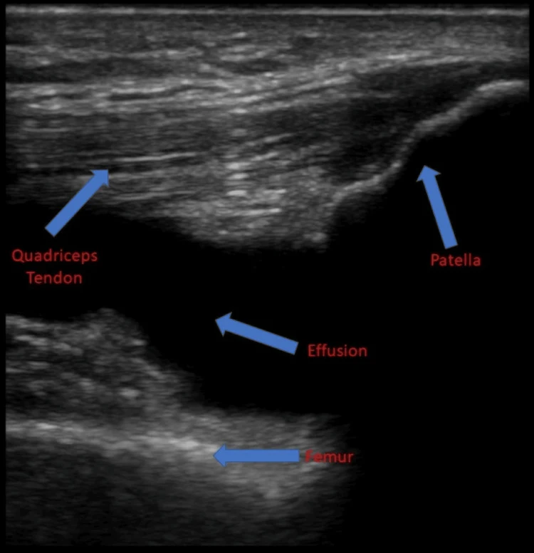
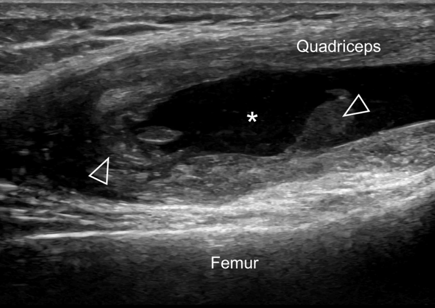
- 線性探頭放在髕骨上緣、股四頭肌腱上,長軸(探頭標記朝頭側):由淺到深依序是股四頭肌腱、膝上囊(suprapatellar recess)、股骨(Schultz 2024)。
- 再轉成短軸,往內外側滑動,找液體最多的一側(多半是外上方)。
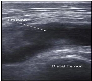
- 膝上囊與關節腔相通,是膝關節積液最先、最明顯出現的地方。
穿刺
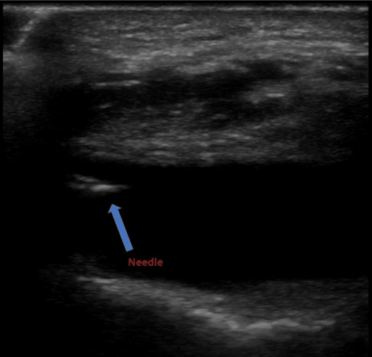
- 短軸下由外側平行探頭進針(in-plane),從髕骨上外緣進入膝上囊,針尖停在液體中央。
- 針越接近水平,越容易全程看到針身;讓針尖遠離股骨表面的軟骨。
- 抽液時用另一手輕壓對側或膝下方,把液體「推」向針尖。
急診實證 急診 RCT 中,超音波並未提高膝關節穿刺的整體成功率,但病人較不痛、總操作時間沒有變長(Wiler 2010)。膝上囊積液大時,觸診法也常成功;超音波的價值在小量積液、肥胖、或不確定是不是關節液的時候。
04髖關節:沿股骨頸的前隱窩
掃描與判讀
- 病人平躺、髖關節中立或微外轉;用凸陣探頭(兒童可用線性),沿股骨頸長軸斜放(矢狀斜切面)。
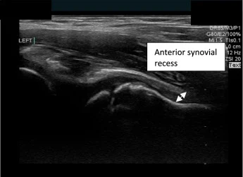
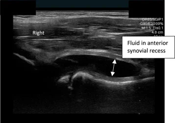
- 畫面中看到股骨頭的圓弧與股骨頸;積液位於股骨頸前表面與髂腰肌後表面之間的前隱窩(Katz-Dana 2024)。
- 兒童的描述性標準:在股骨頸凹處量到的關節囊—滑膜厚度 > 5 mm,或比無症狀側多 2 mm 以上(Tsung 2008)。
- 兒科急診醫師經訓練後可準確辨認:95 位兒童的前瞻研究,敏感度 89.8%、特異度 91.3%(Katz-Dana 2024);較早的小型研究,症狀側敏感度 85%、特異度 93%(Vieira 2010)。
穿刺
- 沿股骨頸長軸、由遠端往近端 in-plane 進針,目標是股骨頭頸交界處前方的液體(原則)。
- 急診醫師在床邊以超音波導引穿刺髖關節已有病例系列描述(Freeman 2007);屍體模型中住院醫師用超音波辨認積液的敏感度 86%、特異度 90%(Berona 2017)。
股神經血管束 股動脈、股靜脈與股神經就在髖關節前方的內側。進針前先用都卜勒把股血管找出來,路徑保持在血管外側。
積液 ≠ 化膿 超音波無法安全地區分兒童化膿性髖關節炎與暫時性滑膜炎(Zamzam 2006);有積液又懷疑感染時,要結合臨床、檢驗,必要時穿刺。
05肩關節:後側盂肱關節入路
- 病人坐姿或側躺,患側手放在對側肩上(內轉內收),讓後關節囊張開。
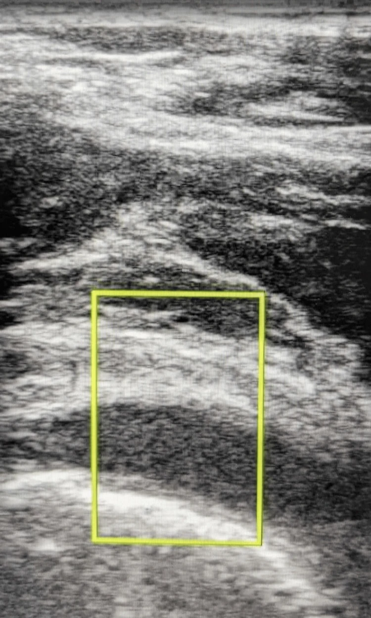
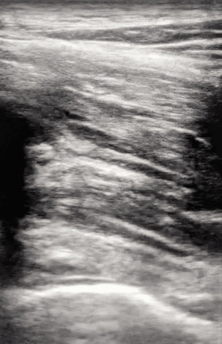
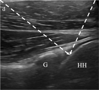
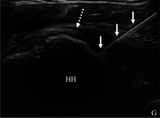
- 探頭橫放在肩胛棘下方、與棘下肌平行,向外滑到肱骨頭與關節盂(glenoid)同時入鏡;後關節隱窩位在兩者之間、棘下肌腱深處(Patel 2023)。
- 可由內往外或由外往內 in-plane 進針,針尖抵達肱骨頭表面、棘下肌腱深層(Patel 2023)。
後側入路的好處 後方沒有大血管神經擋路,肥胖病人也較容易從後方找到關節。前側入路則要注意喙突內側的神經血管,且旁邊有肱二頭肌長頭腱(原則)。
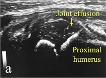
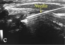
兒童化膿性關節炎 一項兒童化膿性關節炎研究中,作者以超音波導引穿刺肩、踝等關節,圖中可見肩關節積液與穿刺針(Michaud 2024)。
以下內容需要免費訂閱
病例影像判讀與練習題開放給訂閱者。留下電子郵件即可立即閱讀全部內容;同一個瀏覽器一年內不必再輸入。換手機或電腦時,輸入同一個信箱即可。
不收費、不寄廣告。若想收到新章節通知,我們會另寄一封確認信,點了才會開始寄。詳見隱私說明。
10參考文獻與延伸閱讀
- Balint PV, Kane D, Hunter J, et al. Ultrasound guided versus conventional joint and soft tissue fluid aspiration in rheumatology practice: a pilot study. J Rheumatol. 2002;29(10):2209-2213.
- Wiler JL, Costantino TG, Filippone L, Satz W. Comparison of ultrasound-guided and standard landmark techniques for knee arthrocentesis. J Emerg Med. 2010;39(1):76-82. doi:10.1016/j.jemermed.2008.05.012
- Sibbitt WL Jr, Kettwich LG, Band PA, et al. Does ultrasound guidance improve the outcomes of arthrocentesis and corticosteroid injection of the knee? Scand J Rheumatol. 2012;41(1):66-72. doi:10.3109/03009742.2011.599071
- Deng X, Li Y, Li D. Ultrasound-guided versus blind arthrocentesis in knee osteoarthritis: a systematic review and meta-analysis. Medicine (Baltimore). 2025;104(5):e41389. doi:10.1097/MD.0000000000041389
- Adhikari S, Blaivas M. Utility of bedside sonography to distinguish soft tissue abnormalities from joint effusions in the emergency department. J Ultrasound Med. 2010;29(4):519-526. doi:10.7863/jum.2010.29.4.519
- Gottlieb M, Holladay D, Rice M. Current approach to the evaluation and management of septic arthritis. Pediatr Emerg Care. 2019;35(7):509-513. doi:10.1097/PEC.0000000000001874
- Freeman K, Dewitz A, Baker WE. Ultrasound-guided hip arthrocentesis in the ED. Am J Emerg Med. 2007;25(1):80-86. doi:10.1016/j.ajem.2006.08.002
- Tsung JW, Blaivas M. Emergency department diagnosis of pediatric hip effusion and guided arthrocentesis using point-of-care ultrasound. J Emerg Med. 2008;35(4):393-399. doi:10.1016/j.jemermed.2007.10.054
- Vieira RL, Levy JA. Bedside ultrasonography to identify hip effusions in pediatric patients. Ann Emerg Med. 2010;55(3):284-289. doi:10.1016/j.annemergmed.2009.06.527
- Katz-Dana H, Stackievicz R, Dana E, et al. Diagnostic accuracy of point-of-care ultrasound (PoCUS) for the diagnosis of hip effusion in the pediatric emergency department. CJEM. 2024;26(12):875-882. doi:10.1007/s43678-024-00788-z
- Zamzam MM. The role of ultrasound in differentiating septic arthritis from transient synovitis of the hip in children. J Pediatr Orthop B. 2006;15(6):418-422. doi:10.1097/01.bpb.0000228388.32184.7f
- Berona K, Abdi A, Menchine M, et al. Success of ultrasound-guided versus landmark-guided arthrocentesis of hip, ankle, and wrist in a cadaver model. Am J Emerg Med. 2017;35(2):240-244. doi:10.1016/j.ajem.2016.10.056
- Patel RP, McGill K, Motamedi D, et al. Ultrasound-guided interventions of the upper extremity joints. Skeletal Radiol. 2023;52(5):897-909. doi:10.1007/s00256-022-04148-9
圖片來源與授權
- 以下論文的多格拼圖在本頁拆成單格裁切呈現,圖說為本站中文改寫;本頁未使用含病人或志願者身體照片的分格。
- Schultz KL, Johnson K, Grenz PM, et al. Increasing efficiency with point-of-care ultrasound-guided arthrocentesis: a case report and procedure demonstration. Cureus 2024;16(6):e62082, doi:10.7759/cureus.62082(Figure 2、3),CC BY 4.0。
- Vaid Z, Shah AB, Sheth D, et al. Melioidosis presenting as a solitary liver abscess with culture-negative septic arthritis: a case report. Cureus 2026;18(9):e116326, doi:10.7759/cureus.116326(Figure 2),CC BY 4.0。
- Halupa AJ, Strony RJ, Bulbin DH, et al. Pseudogout diagnosed by point-of-care ultrasound. Clin Pract Cases Emerg Med 2019;3(4):425-427, doi:10.5811/cpcem.2019.7.43244(Image 1、3),CC BY 4.0。
- Katz-Dana H, Stackievicz R, Dana E, et al. Diagnostic accuracy of point-of-care ultrasound (PoCUS) for the diagnosis of hip effusion in the pediatric emergency department. CJEM 2024;26(12):875-882, doi:10.1007/s43678-024-00788-z(Fig. 1),CC BY 4.0。
- Storch ID, Nippert J. A pain in the hip: sexually transmitted septic arthritis diagnosed with bedside ultrasound in the emergency department. Cureus 2025;17(12):e98653, doi:10.7759/cureus.98653(Figure 1、2),CC BY 4.0。
- Michaud J, Dutron S, Pico J, et al. The feasibility and safety of ultrasound-guided puncture for treatment of septic arthritis in children. Ital J Pediatr 2024;50(1):198, doi:10.1186/s13052-024-01746-2(Fig. 1、2),CC BY 4.0。
- Patel RP, McGill K, Motamedi D, et al. Ultrasound-guided interventions of the upper extremity joints. Skeletal Radiol 2023;52(5):897-909, doi:10.1007/s00256-022-04148-9(Fig. 1d、2、8d),CC BY 4.0。
- Patitsas P, Davis R, Strony R. Point-of-care ultrasound-directed evaluation of elbow effusion. Clin Pract Cases Emerg Med 2019;3(3):286-288, doi:10.5811/cpcem.2019.5.41674(Image 1–3),CC BY 4.0。
- Mogami R, Vaz JLP, Chagas YFB, et al. Ultrasound of ankles in the diagnosis of complications of chikungunya fever. Radiol Bras 2017;50(2):71-75, doi:10.1590/0100-3984.2016.0221(Figure 1),CC BY 4.0。
- Shmerling A, Bravman JT, Khodaee M. Morel-Lavallée lesion of the knee in a recreational frisbee player. Case Rep Orthop 2016;2016:8723489, doi:10.1155/2016/8723489(Figure 3a、3d),CC BY 4.0。
- Yamada T. When joint aspiration fails: point-of-care ultrasound for diagnosing chronic gout in prolonged monoarthritis. J Gen Fam Med 2026;27(5):e70155, doi:10.1002/jgf2.70155(Figure 1A),CC BY 4.0。