超音波圖譜 › 第 27 章 · 超音波導引處置
27.5 膿瘍引流與軟組織異物
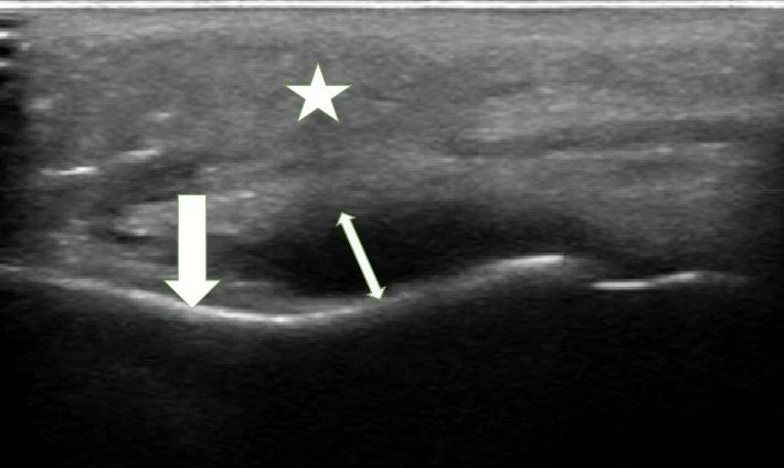
這一章的重點 紅腫熱痛不一定有膿。超音波回答三個問題:有沒有可以引流的積液、積液旁邊有沒有血管、要從哪裡、多深下刀。異物則看亮線、後方陰影或彗星尾、周圍低回音暈環,並沿異物長軸切開。
01使用時機與證據
進針、無菌與 in-plane 等共通原則見本章母頁:超音波導引處置。
什麼時候拿探頭
- 蜂窩性組織炎還是膿瘍?理學檢查摸不出波動感、或範圍大到不知道從哪裡切時。
- 切之前:確認積液位置、大小與深度,並用彩色都卜勒排除血管、假性動脈瘤或淋巴結。
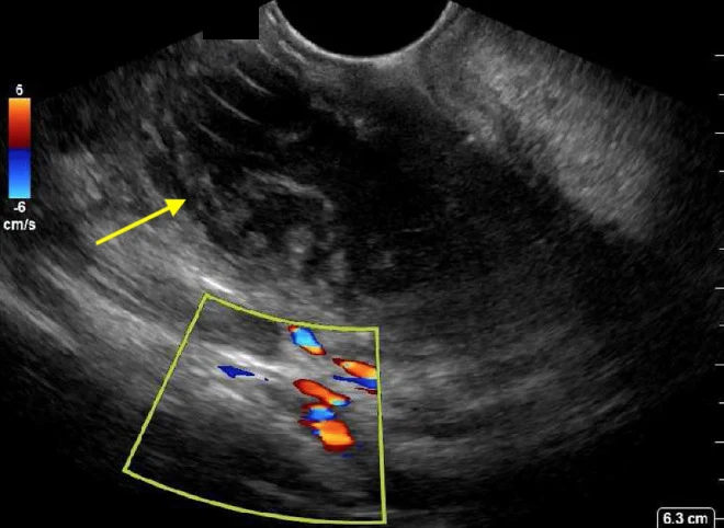
- 扁桃腺周圍膿瘍:口內超音波區分膿瘍與蜂窩性組織炎,並看清頸動脈的位置。
- 傷口疑似殘留異物:尤其木頭、植物等 X 光看不到的材質。
- 引流後追蹤:殘留的腔室、治療失敗回診時再評估。
診斷準確度 14 篇前瞻研究、2,656 位病人的統合分析:床邊超音波(POCUS)診斷膿瘍的敏感度 94.6%、特異度 85.4%;有 10.3% 的病人因超音波而正確改變處置,0.7% 錯誤改變(Gottlieb 2020)。另兩篇統合分析結果相近:敏感度 97%/特異度 83%(Subramaniam 2016)、96.2%/82.9%(Barbic 2017)。
兒童較難 兒童次族群敏感度 89.9%、特異度 79.9%,低於成人的 98.7%、91.0%(Gottlieb 2020)。兒童專屬統合分析:超音波敏感度 0.90、特異度 0.80,理學檢查 0.84、0.69(Wu 2022)。
在臨床判斷為蜂窩性組織炎、看不出明顯膿瘍的成人中,軟組織超音波改變了 71/126(56%)的處置:找到需要引流的隱匿膿瘍,也讓一部分人免於不必要的切開(Tayal 2006)。
02掃描技巧
- 高頻線性探頭,深度先調淺,把焦點放在皮下層;很表淺的病灶(手指、指甲周圍)可用水浴或厚凝膠墊增加距離。
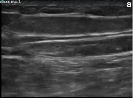
- 先掃健側或周邊正常皮膚,記住這個人皮膚、皮下脂肪、筋膜、肌肉的正常樣子。
- 兩個垂直切面(橫切、縱切)扇掃整個紅腫範圍,量出積液的長、寬、深與皮膚到積液的距離。
- 輕壓探頭:膿液內的碎屑會流動或漩動(swirl),有助於分辨等回音的膿與實質組織。
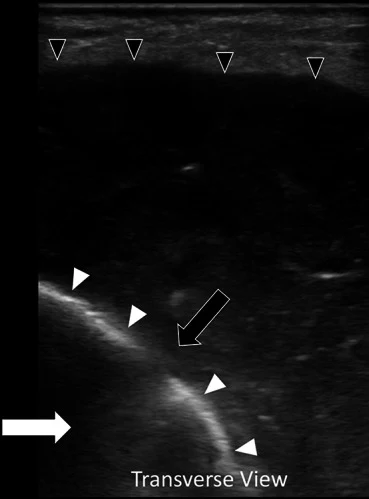
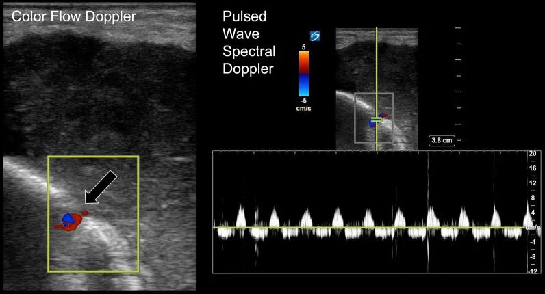
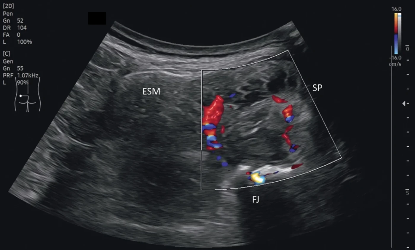
- 彩色都卜勒:積液中央不該有血流;周邊充血常見。中央有血流或搏動,先停手再判斷。
| 蜂窩性組織炎 | 膿瘍 | |
|---|---|---|
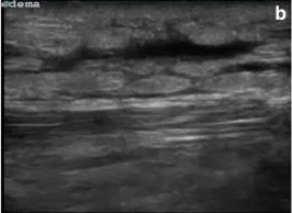
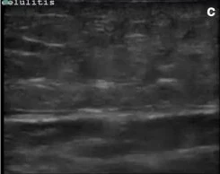
| 皮下脂肪 | 回音變亮、層次模糊;水腫進入脂肪小葉間,形成鵝卵石(cobblestone)樣 | 周邊組織常同樣發炎變亮 |
| 積液 | 無局部積液,只有網狀低回音條紋 | 局部無回音至混合回音的積液,邊界可不規則 |
| 後方 | — | 常見後方回音增強 |
| 壓迫 | — | 內容物流動、漩動 |
| 都卜勒 | 整片充血 | 周邊充血、中央無血流 |
鵝卵石樣不等於膿瘍:水腫(心衰竭、低白蛋白、淋巴水腫)也會出現,要配合臨床。
03切開引流與針吸
兩種用法
- 定位(static):掃描後在皮膚標出積液最大、最淺的位置與方向,記下深度,再照一般方式切開引流。大部分皮下膿瘍這樣就夠。
- 即時導引(dynamic):針吸或放置引流時探頭留在現場,in-plane 看著針尖進入積液。適合較深、靠近血管神經,或位置特殊(臉、手、乳房、扁桃腺周圍)的膿瘍。
- 引流後再掃一次:看還有沒有沒打開的腔室(loculation)。
切之前三件事 ①彩色都卜勒排除血管、假性動脈瘤(尤其鼠蹊部、注射藥物使用者、近期導管處置處)與腫大淋巴結;②看清楚積液與深部構造(骨頭、關節、肌腱鞘、體腔)的關係;③量好皮膚到積液的深度,決定刀與針的長度。
積液多大才值得切、多大可以只用抗生素,目前沒有一致的切點;本頁不給數字,請依積液大小、深度、臨床病程與各院規範判斷。
04扁桃腺周圍膿瘍與頭頸部
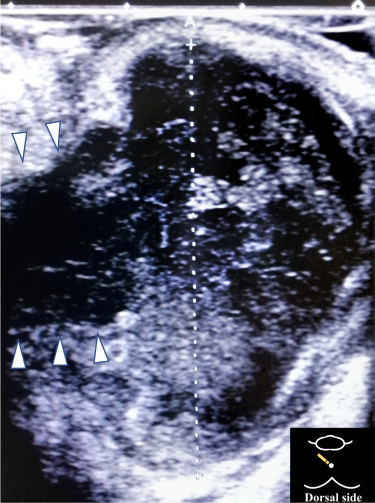
- 口內法:腔內(endocavitary)探頭套上探頭套,直接放在腫脹的扁桃腺上;經頸法則從下頷角下方掃。
- 找扁桃腺內或旁邊的低回音至混合回音積液,並用彩色都卜勒找出頸內動脈的位置與距離,再決定針吸的方向與深度。
- 隨機試驗(28 位成人):口內超音波組正確診斷率 100%,傳統目視針吸組 64%;超音波組耳鼻喉科會診 7% vs 50%,電腦斷層 0% vs 35%(Costantino 2012)。
準確度不是完美 18 篇研究的統合分析:超音波診斷扁桃腺周圍膿瘍的敏感度 86%、特異度 76%;急診醫師自做的 POCUS 敏感度 74%、特異度 79%,口內法敏感度高於經頸法(91% vs 80%)(Kim 2023)。超音波陰性但臨床高度懷疑時,仍需進一步影像或會診。
同一單位在推廣超音波後的回溯比較:急診醫師針吸成功率 POCUS 組 89.1%、未用超音波組 24.5%(Gibbons 2020,回溯性研究)。
以下內容需要免費訂閱
病例影像判讀與練習題開放給訂閱者。留下電子郵件即可立即閱讀全部內容;同一個瀏覽器一年內不必再輸入。換手機或電腦時,輸入同一個信箱即可。
不收費、不寄廣告。若想收到新章節通知,我們會另寄一封確認信,點了才會開始寄。詳見隱私說明。
08參考文獻與延伸閱讀
- Gottlieb M, Avila J, Chottiner M, Peksa GD. Point-of-care ultrasonography for the diagnosis of skin and soft tissue abscesses: a systematic review and meta-analysis. Ann Emerg Med. 2020;76(1):67-77. doi:10.1016/j.annemergmed.2020.01.004
- Subramaniam S, Bober J, Chao J, Zehtabchi S. Point-of-care ultrasound for diagnosis of abscess in skin and soft tissue infections. Acad Emerg Med. 2016;23(11):1298-1306. doi:10.1111/acem.13049
- Barbic D, Chenkin J, Cho DD, Jelic T, Scheuermeyer FX. In patients presenting to the emergency department with skin and soft tissue infections what is the diagnostic accuracy of point-of-care ultrasonography for the diagnosis of abscess compared to the current standard of care? A systematic review and meta-analysis. BMJ Open. 2017;7(1):e013688. doi:10.1136/bmjopen-2016-013688
- Wu J, Ge L, Wang X, Jin Y. Role of point-of-care ultrasound (POCUS) in the diagnosis of an abscess in paediatric skin and soft tissue infections: a systematic review and meta-analysis. Med Ultrason. 2022;24(3):339-347. doi:10.11152/mu-3166
- Tayal VS, Hasan N, Norton HJ, Tomaszewski CA. The effect of soft-tissue ultrasound on the management of cellulitis in the emergency department. Acad Emerg Med. 2006;13(4):384-388. doi:10.1197/j.aem.2005.11.074
- Costantino TG, Satz WA, Dehnkamp W, Goett H. Randomized trial comparing intraoral ultrasound to landmark-based needle aspiration in patients with suspected peritonsillar abscess. Acad Emerg Med. 2012;19(6):626-631. doi:10.1111/j.1553-2712.2012.01380.x
- Kim DJ, Burton JE, Hammad A, et al. Test characteristics of ultrasound for the diagnosis of peritonsillar abscess: a systematic review and meta-analysis. Acad Emerg Med. 2023;30(8):859-869. doi:10.1111/acem.14660
- Gibbons RC, Costantino TG. Evidence-based medicine improves the emergent management of peritonsillar abscesses using point-of-care ultrasound. J Emerg Med. 2020;59(5):693-698. doi:10.1016/j.jemermed.2020.06.030
- Davis J, Czerniski B, Au A, et al. Diagnostic accuracy of ultrasonography in retained soft tissue foreign bodies: a systematic review and meta-analysis. Acad Emerg Med. 2015;22(7):777-787. doi:10.1111/acem.12714
- Mohammadi A, Ghasemi-Rad M, Khodabakhsh M. Non-opaque soft tissue foreign body: sonographic findings. BMC Med Imaging. 2011;11:9. doi:10.1186/1471-2342-11-9
- Lulla A, Whitman T, Amii R, Chiem AT. Role of ultrasound in the identification of longitudinal axis in soft-tissue foreign body extraction. West J Emerg Med. 2016;17(6):819-821. doi:10.5811/westjem.2016.8.30988
- Shelhoss SC, Burgin CM. Maximizing foreign body detection by ultrasound with the water bath technique coupled with the focal zone advantage: a technical report. Cureus. 2022;14(11):e31577. doi:10.7759/cureus.31577
圖片來源與授權
- 以下論文的多格拼圖在本頁拆成單格裁切呈現,圖說為本站中文改寫;部分影像上的超音波儀器廠牌字樣與標誌已以色塊遮蔽或裁除。
- Magalhães L, Martins SRP, Nogué R. The role of point-of-care ultrasound in the diagnosis and management of necrotizing soft tissue infections. Ultrasound J 2020;12:3, doi:10.1186/s13089-020-0153-4(Fig. 5),CC BY 4.0。
- Hori H, Sugawara H. Application of point-of-care ultrasound scanning for the diagnosis of perianal abscess. Cureus 2022;14(1):e21622, doi:10.7759/cureus.21622(Fig. 2),CC BY 3.0。
- Werenski H, Ford K, Casey D, et al. Abscess pulsatility: a sonographic sign of osteomyelitis. Ultrasound J 2023;15:41, doi:10.1186/s13089-023-00339-0(Fig. 3、4),CC BY 4.0。
- Schöll E, Mair AC, Litz RJ. Sonographic detection of a lumbar facet joint abscess with epidural involvement. Ultrasound Int Open 2026;12:a29725876, doi:10.1055/a-2972-5876(Fig. 4),CC BY 4.0。
- Barrett J, Moore C, Kramer JA, et al. You've gotten under my skin: how to make a simple, non-perishable, low-cost soft tissue infection ultrasound simulator. POCUS J 2025;10(1):119-122, doi:10.24908/pocusj.v10i01.18407(Fig. 6),CC BY 4.0。
- Floyd J, Dahl B, Whited M, et al. Peritonsillar abscess and post-aspiration bleed identified with point-of-care ultrasound using endocavitary probe: a case report. Clin Pract Cases Emerg Med 2024;8(2):129-132, doi:10.5811/cpcem.1645(Image),CC BY 4.0。
- Scheier E, Gold B. Dental abscess on POCUS: a retrospective case series analysis. POCUS J 2026;11(1):72-76, doi:10.24908/pocusj.v11i01.19441(Fig. 2),CC BY 4.0。
- Mohammadi A, Ghasemi-Rad M, Khodabakhsh M. Non-opaque soft tissue foreign body: sonographic findings. BMC Med Imaging 2011;11:9, doi:10.1186/1471-2342-11-9(Fig. 1–4),CC BY 2.0。
- Lulla A, Whitman T, Amii R, Chiem AT. Role of ultrasound in the identification of longitudinal axis in soft-tissue foreign body extraction. West J Emerg Med 2016;17(6):819-821, doi:10.5811/westjem.2016.8.30988(Fig. 1、2),CC BY 4.0。
- Eswine SL, Chassee TP, Benner CA, et al. Lost in the woods: a case report of a radiolucent wooden foreign body in a child after negative CT, detected on repeat ultrasound. Cureus 2026;18(7):e112961, doi:10.7759/cureus.112961(Fig. 2、3),CC BY 4.0。
- Shelhoss SC, Burgin CM. Maximizing foreign body detection by ultrasound with the water bath technique coupled with the focal zone advantage: a technical report. Cureus 2022;14(11):e31577, doi:10.7759/cureus.31577(Fig. 3A、3B),CC BY 3.0。
- McCreary D, Sarvesh B, Munir M. What lies beneath? Using point of care ultrasound (POCUS) to identify soft tissue foreign bodies in children and adults: a literature review. POCUS J 2025;10(1):110-118, doi:10.24908/pocusj.v10i01.18072(Fig. 5、6),CC BY 4.0。