超音波圖譜 › 第 21 章 · 肌肉骨骼
21.2 關節積液與脫位:髖、膝、肘、肩
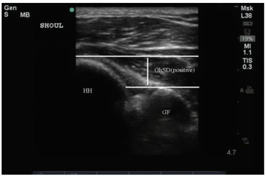
這一章的重點 關節積液在超音波上就是關節隱窩被無回音或低回音液體撐開,和健側比最可靠。超音波可以回答「有沒有積液、在哪裡最好抽」,但不能分辨化膿、痛風或出血,懷疑化膿性關節炎還是要抽。肩脫位用後側切面看肱骨頭和關節盂的相對位置,復位後馬上再看一次。
01使用時機:關節腫痛的第一個問題
共通基本功請先看母頁:肌肉骨骼超音波:基本功與正常構造。關節穿刺的步驟在第 27 章超音波導引處置,本頁只講定位。
適應症(本課程講義)
- 急性或慢性關節腫脹;膝蓋有浮髕感(ballotable patella)。
- 鑑別:痛風、假性痛風、類風濕性關節炎、蜂窩性組織炎、髕前滑囊炎、膿瘍。
- 關節積液的原因:感染性發炎、非感染性發炎、腫瘤、外傷。
- 兒童跛行、拒絕負重:找髖關節積液。
超音波改變了什麼
急診 54 位關節腫痛病人中,床邊超音波找到積液的有 40.7%;超音波改變了 65% 病人的處置。原本計畫做關節穿刺的 39 位,看完超音波後只有 20 位真的去抽;這項研究的目的正是區分軟組織異常與關節積液(Adhikari 2010)。
超音波不能做的事 積液的回音高低不能區分化膿性、結晶性或出血性。懷疑化膿性關節炎時,即使積液看起來是乾淨的無回音,仍要抽出關節液送檢。
02髖關節:前側隱窩
掃法
- 病人仰臥,髖關節稍外展、外旋。
- 探頭平行股骨頸斜放(講義口訣:約 45 度、朝肚臍方向);兒童用線性探頭,成人或體型大改用凸陣探頭。
- 畫面上由內而外依序是:髖臼、股骨頭、股骨頸,上方是髂腰肌。
- 前側滑膜隱窩在股骨頸前緣和髂腰肌後緣之間,量它的厚度,並和健側比較。
研究使用的陽性標準:前側隱窩大於 5 mm,或比對側多 2 mm 以上(Katz-Dana 2024,引用既有文獻的定義)。
準確度(兒童)
- 兒科急診醫師受訓後,症狀側髖關節敏感度 85%、特異度 93%(28 位病人、55 個髖關節;Vieira 2010)。
- 單中心前瞻研究 95 位病人:敏感度 89.8%、特異度 91.3%,與放射科超音波一致性 κ = 0.81(Katz-Dana 2024)。
- 統合分析 4 篇、526 個髖關節:合併敏感度 88%、特異度 97%(Gravel 2026)。
有積液之後 超音波只能說「有積液」,不能分辨暫時性滑膜炎與化膿性關節炎。要結合發燒、能否負重、發炎指數等臨床資料,依各院流程決定是否穿刺或會診。
03膝與肘:髕上隱窩與鷹嘴窩
膝:髕上隱窩(本課程講義)
- 機器放在操作者視線正前方;膝蓋微彎(可墊毛巾)。
- 線性探頭(講義寫 5–10 MHz)縱向放在髕骨上緣。
- 依序認出髕骨、股骨、股四頭肌腱與脂肪墊。
- 在股四頭肌腱深處找無回音的液體,並和另一腳比較。
- 要抽的話:局部麻醉後,由外往內以 in-plane 進針,避開肌腱、骨頭與韌帶(步驟見第 27 章)。
肘:後側鷹嘴窩
- 肘彎曲,探頭縱向放在三頭肌腱上、一端頂在鷹嘴;三頭肌腱下方看到的液體就在關節內(Agrait Gonzalez 2025)。
- 轉成短軸,可以看到積液在鷹嘴窩內,並作為 in-plane 穿刺的切面。
- 肘關節外的疾病(鷹嘴滑囊炎、上髁炎)不會造成被動與主動伸直受限,可以幫忙和關節內積液區分(Agrait Gonzalez 2025)。
超音波導引穿刺的證據
膝關節穿刺的急診隨機試驗:成功率與傳統觸診法沒有差別(37/39 vs 25/27),但病人疼痛較低、操作者覺得較容易(Wiler 2010)。大體模型中,18 位住院醫師用超音波找髖、踝、腕積液的敏感度 86%、特異度 90%,穿刺成功率則沒有顯著差異(Berona 2017)。
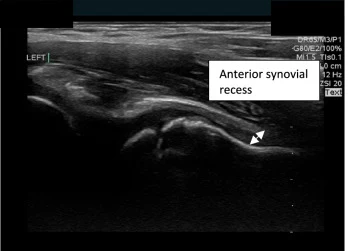
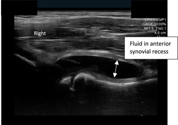
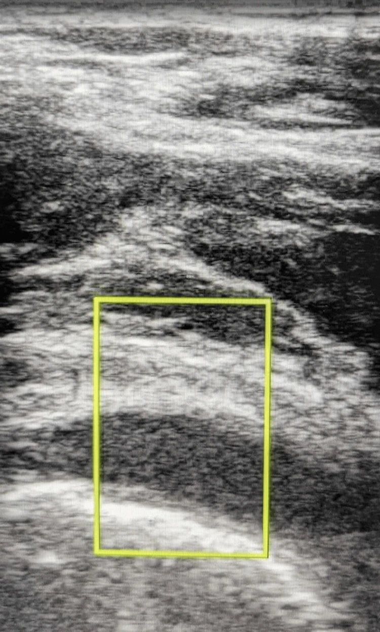
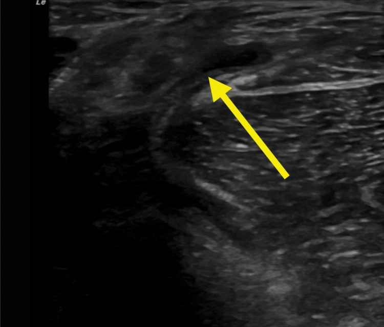
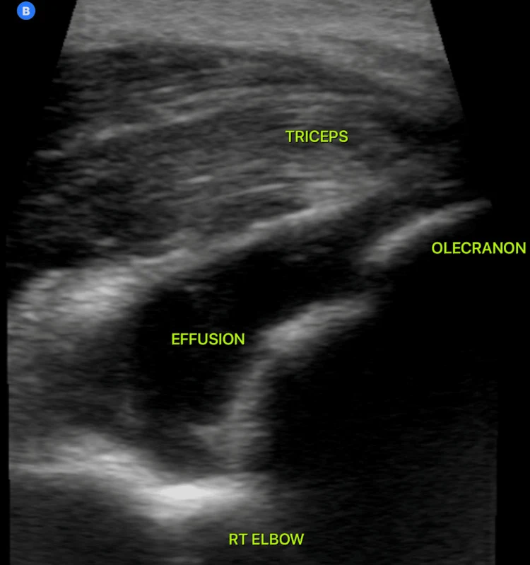
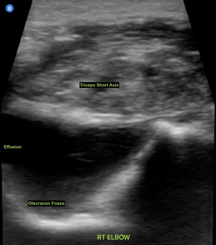
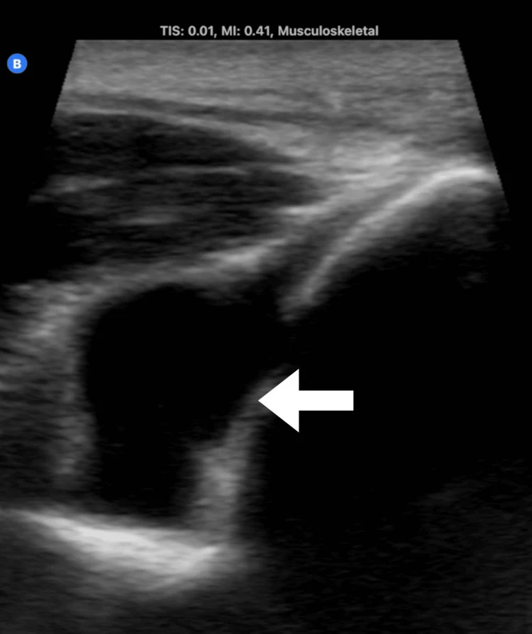
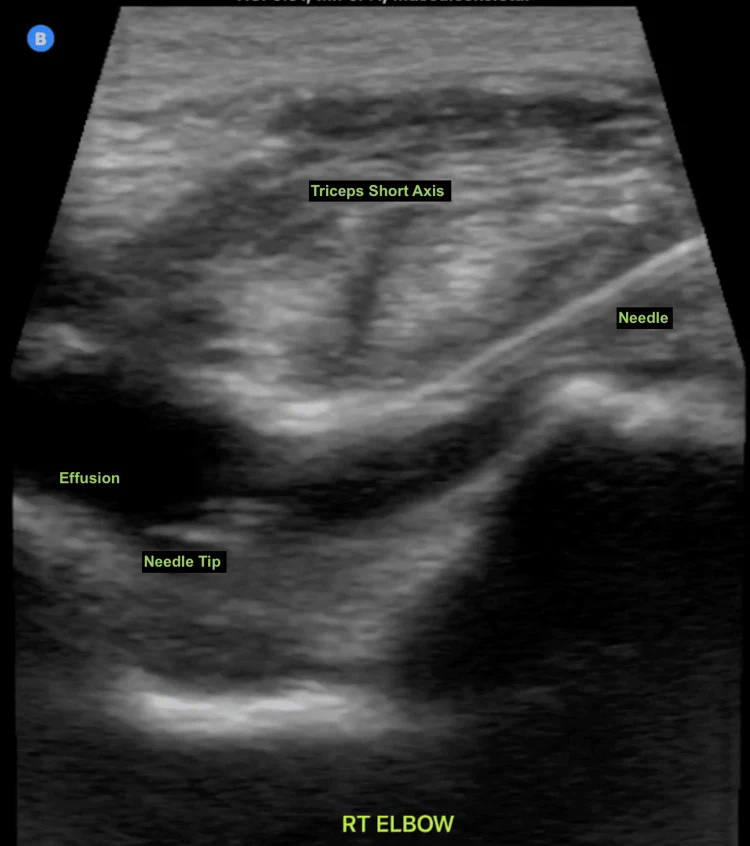
髖關節積液兩張為同一研究的正常與異常示例;成人髖關節與肘關節影像來自不同個案報告。
04肩與胸鎖關節:後側切面找積液
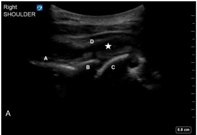
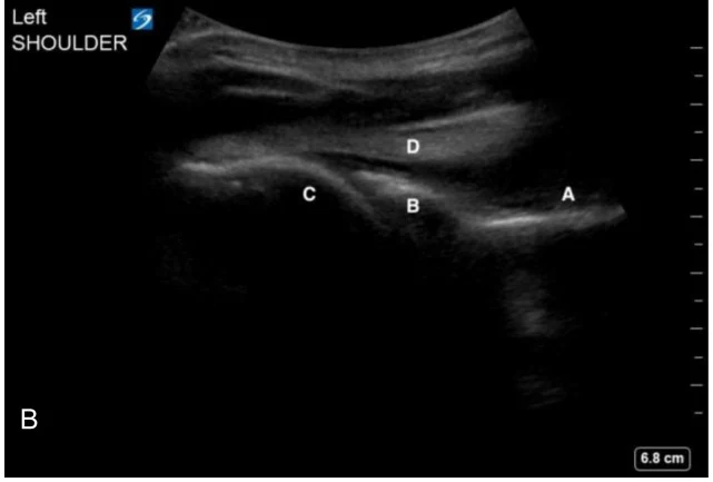
肩關節積液在後側最容易看:探頭橫放在肩後側,可以看到肩胛棘、關節盂、肱骨頭與棘下肌腱。積液或積血會出現在棘下肌腱深處、肱骨頭與關節盂之間(Padrez 2022)。
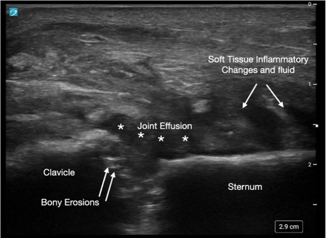
胸鎖關節表淺,用線性探頭橫放在鎖骨內側端與胸骨之間即可;看關節囊是否撐開、骨頭表面是否有侵蝕。原文個案為化膿性胸鎖關節炎,同時有骨侵蝕與周邊軟組織發炎(Koshy 2024)。
以下內容需要免費訂閱
病例影像判讀與練習題開放給訂閱者。留下電子郵件即可立即閱讀全部內容;同一個瀏覽器一年內不必再輸入。換手機或電腦時,輸入同一個信箱即可。
不收費、不寄廣告。若想收到新章節通知,我們會另寄一封確認信,點了才會開始寄。詳見隱私說明。
08參考文獻與延伸閱讀
- Adhikari S, Blaivas M. Utility of bedside sonography to distinguish soft tissue abnormalities from joint effusions in the emergency department. J Ultrasound Med. 2010;29(4):519-26. doi:10.7863/jum.2010.29.4.519
- Vieira RL, Levy JA. Bedside ultrasonography to identify hip effusions in pediatric patients. Ann Emerg Med. 2010;55(3):284-9. doi:10.1016/j.annemergmed.2009.06.527
- Katz-Dana H, Stackievicz R, Dana E, et al. Diagnostic accuracy of point-of-care ultrasound (PoCUS) for the diagnosis of hip effusion in the pediatric emergency department. CJEM. 2024;26(12):875-882. doi:10.1007/s43678-024-00788-z
- Gravel CA, Neal JT, Dodderer J, et al. Point-of-care ultrasound for hip effusion in children: A systematic review and meta-analysis. Am J Emerg Med. 2026;102:148-154. doi:10.1016/j.ajem.2026.01.034
- Wiler JL, Costantino TG, Filippone L, et al. Comparison of ultrasound-guided and standard landmark techniques for knee arthrocentesis. J Emerg Med. 2010;39(1):76-82. doi:10.1016/j.jemermed.2008.05.012
- Berona K, Abdi A, Menchine M, et al. Success of ultrasound-guided versus landmark-guided arthrocentesis of hip, ankle, and wrist in a cadaver model. Am J Emerg Med. 2017;35(2):240-244. doi:10.1016/j.ajem.2016.10.056
- Gottlieb M, Patel D, Marks A, et al. Ultrasound for the diagnosis of shoulder dislocation and reduction: A systematic review and meta-analysis. Acad Emerg Med. 2022;29(8):999-1007. doi:10.1111/acem.14454
- Secko MA, Reardon L, Gottlieb M, et al. Musculoskeletal Ultrasonography to Diagnose Dislocated Shoulders: A Prospective Cohort. Ann Emerg Med. 2020;76(2):119-128. doi:10.1016/j.annemergmed.2020.01.008
- Lahham S, Becker B, Chiem A, et al. Pilot Study to Determine Accuracy of Posterior Approach Ultrasound for Shoulder Dislocation by Novice Sonographers. West J Emerg Med. 2016;17(3):377-82. doi:10.5811/westjem.2016.2.29290
- Agrait Gonzalez MF, Mercado A, Ortiz Diaz S. A Different Type of Tennis Elbow: Spontaneous Septic Arthritis of the Elbow in a Previously Healthy 40-Year-Old Male. Cureus. 2025;17(7):e87228. doi:10.7759/cureus.87228
- Padrez KA, Shyy W, Gandhi K, et al. Identification of Spontaneous Shoulder Hemarthrosis with Point-of-Care Ultrasound in the Emergency Department. Clin Pract Cases Emerg Med. 2022;6(2):177-179. doi:10.5811/cpcem.2021.7.53408
圖片來源與授權
- 以下論文的多格拼圖在本頁拆成單格裁切呈現,圖說為本站中文改寫。
- Katz-Dana H, Stackievicz R, Dana E, et al. Diagnostic accuracy of point-of-care ultrasound (PoCUS) for the diagnosis of hip effusion in the pediatric emergency department. CJEM 2024;26(12):875-882, doi:10.1007/s43678-024-00788-z(Fig. 1),CC BY 4.0。
- Storch ID, Nippert J. A Pain in the Hip: Sexually Transmitted Septic Arthritis Diagnosed With Bedside Ultrasound in the Emergency Department. Cureus 2025;17(12):e98653, doi:10.7759/cureus.98653(Fig. 1),CC BY 4.0。
- Anies EP, Waltz RA, Lacey S. A Baker's Cyst With Distal Extramuscular Extension: A Case Report. Cureus 2025;17(1):e77502, doi:10.7759/cureus.77502(Fig. 3),CC BY 4.0。
- Agrait Gonzalez MF, Mercado A, Ortiz Diaz S. A Different Type of Tennis Elbow: Spontaneous Septic Arthritis of the Elbow in a Previously Healthy 40-Year-Old Male. Cureus 2025;17(7):e87228, doi:10.7759/cureus.87228(Fig. 2、Fig. 3、Fig. 4),CC BY 4.0。
- Rodriguez Aquino EG, Agrait Gonzalez MF, Marrero Medina S, et al. A Rare Case of Spontaneous Elbow Osteomyelitis Presenting With Aseptic Effusion. Cureus 2025;17(3):e80305, doi:10.7759/cureus.80305(Fig. 2),CC BY 4.0。
- Padrez KA, Shyy W, Gandhi K, et al. Identification of Spontaneous Shoulder Hemarthrosis with Point-of-Care Ultrasound in the Emergency Department. Clin Pract Cases Emerg Med 2022;6(2):177-179, doi:10.5811/cpcem.2021.7.53408(Image 2),CC BY 4.0。
- Koshy GV, Davis R, Wilson C. Septic Arthritis of the Sternoclavicular Joint. Clin Pract Cases Emerg Med 2024;8(1):60-63, doi:10.5811/cpcem.1563(Image 2),CC BY 4.0。
- Lahham S, Becker B, Chiem A, et al. Pilot Study to Determine Accuracy of Posterior Approach Ultrasound for Shoulder Dislocation by Novice Sonographers. West J Emerg Med 2016;17(3):377-82, doi:10.5811/westjem.2016.2.29290(Fig. 4、Fig. 3),CC BY 4.0。
- Gayda S, Kohen B, Boccio E. Woman With Shoulder Pain: Posterior Shoulder Dislocation Diagnosed With Point-of-Care Ultrasound. Cureus 2024;16(7):e64180, doi:10.7759/cureus.64180(Fig. 1),CC BY 4.0。
- Lahr R, Wheeler F, Espinosa J, et al. Use of Handheld Bedside Ultrasound to Confirm Successful Reduction of an Anterior Shoulder Dislocation. Cureus 2024;16(1):e52089, doi:10.7759/cureus.52089(Fig. 2、Fig. 3),CC BY 4.0。