超音波圖譜 › 第 18 章 · 婦科
婦科超音波(Gynecology)
這一章的重點 急診的婦科超音波先驗孕,再回答三件事:骨盆腔有沒有游離液、附件有沒有腫塊、子宮有沒有問題。卵巢扭轉要靠腫大的卵巢、水腫與漩渦徵象去懷疑,都卜勒有血流不能排除;超音波陰性也不能排除 TOA。
01使用時機:急診的骨盆腔超音波要回答什麼
女性下腹痛、骨盆腔痛或不正常陰道出血,是急診每天都會遇到的主訴。床邊骨盆腔超音波不是要取代婦產科或影像科的正式檢查,而是幫你快速回答幾個會改變處置速度的問題。ACEP 超音波指引把骨盆腔超音波列為急診超音波的應用項目(ACEP 2023)。
床邊要回答的問題
- 有沒有懷孕?先驗孕。懷孕者先看子宮內有沒有妊娠,子宮外孕的流程請見第 16 章 早期妊娠。
- 骨盆腔有沒有游離液?量多、或合併血行動力不穩,要往出血(破裂囊腫、子宮外孕)想。
- 附件(卵巢、輸卵管)有沒有腫塊?單側腫大的卵巢+劇痛,要先排除扭轉。
- 子宮本身:肌瘤、內膜厚度、子宮內避孕器(IUD)位置。
學完這一章,你應該能夠
- 用經腹(transabdominal, TA)與經陰道(transvaginal, TV)掃出子宮、卵巢與子宮直腸凹(pouch of Douglas)。
- 認出出血性囊腫的網狀內部回音與血塊,知道它多半會自己吸收。
- 說出卵巢扭轉的超音波徵象,以及都卜勒有血流也不能排除扭轉的理由。
- 認出輸卵管卵巢膿瘍(tubo-ovarian abscess, TOA)與輸卵管發炎的徵象。
- 判讀停經後出血的內膜厚度、IUD 的位置。
02掃描技巧與正常解剖
經腹(TA):先做、看大範圍
- 凸面探頭放恥骨上方,縱切與橫切各掃一遍。膀胱有尿時,膀胱就是看子宮與卵巢的聲窗。
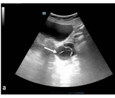
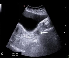
- 縱切找子宮長軸、子宮頸與膀胱,子宮後方就是子宮直腸凹,是骨盆腔游離液最先聚集的位置。
- 橫切從子宮底部往兩側找卵巢,卵巢常在子宮兩側、髂內血管附近;位置個別差異很大。
- 適合:大的腫塊、子宮超出骨盆、無法或不適合經陰道檢查(例如未有性經驗、病人拒絕)。
經陰道(TV):解析度高、看細節
- 腔內探頭頻率較高、距離器官近,卵巢濾泡、內膜、輸卵管看得比經腹清楚;膀胱要排空。
- 探頭套上保護套、潤滑後置入,徵得病人同意並依各院規範安排陪檢人員。
- 矢狀面看子宮與子宮頸,轉 90 度成冠狀面,再往兩側側壁找卵巢。
- 探頭本身可以當「觸診」:壓到哪裡最痛,常就是病灶所在。
兩種掃法互補 經腹先看全貌與游離液、經陰道再看細節。只做經陰道可能漏掉位置高或很大的腫塊;只做經腹則常看不清卵巢內部構造。
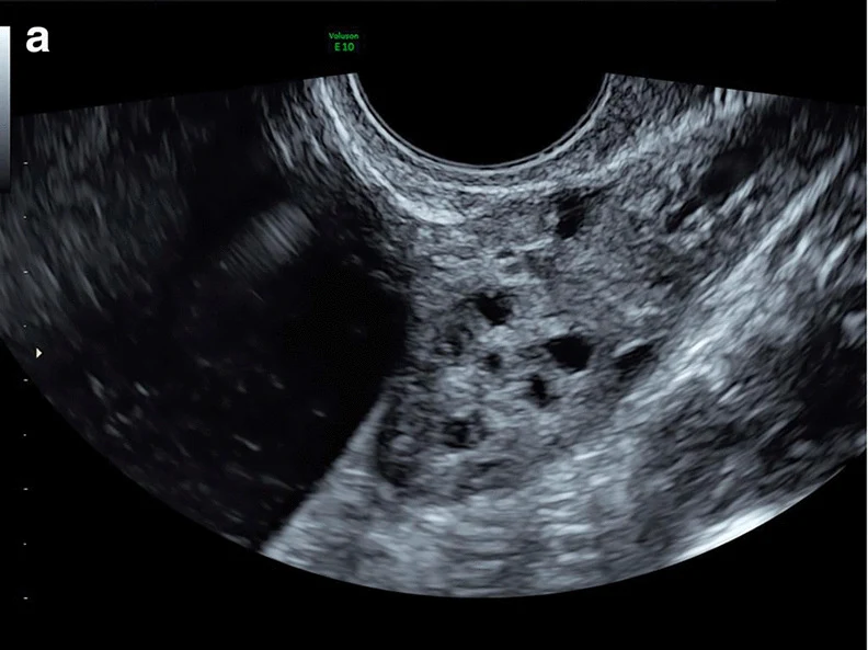
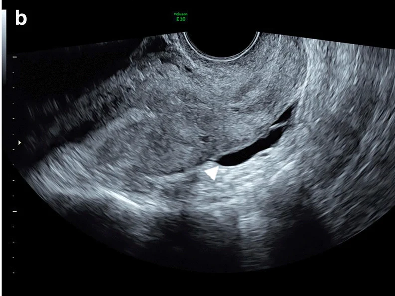
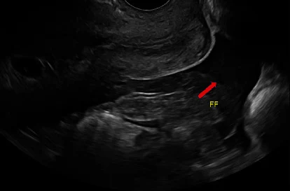
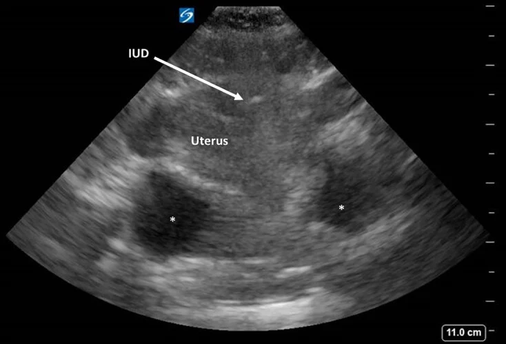
後兩張影像來自懷孕病人,放在這裡只是示範游離液的位置;懷孕病人的判讀請見第 16 章。
03卵巢囊腫:功能性、出血性與破裂
功能性與出血性囊腫
- 生育年齡女性的卵巢常有濾泡或黃體,多數是生理性的。單純囊腫:壁薄、內部無回音、後方回音增強。
- 出血性囊腫:濾泡或黃體內出血。典型外觀是細網狀(蕾絲狀)的纖維蛋白絲,或邊緣內凹、沒有血流的血塊。
- 盲法判讀研究中,約 90% 的出血性囊腫至少有纖維蛋白絲或回縮血塊其中一項;被誤判為出血性囊腫的 2 例,病理是子宮內膜瘤(Patel 2005)。
- 內部網狀構造沒有血流;有血流的實心成分要想到腫瘤。
破裂與後續追蹤
- 囊腫(尤其黃體)破裂可造成腹腔內出血。超音波重點是游離液的量與是否延伸到上腹部(Morison 囊袋),再配合生命徵象與血色素。
- 子宮直腸凹的液體若有回音或有血塊,傾向是血而不是生理性液體。
- 一定要驗孕:懷孕婦女的附件腫塊合併腹內出血,先當子宮外孕處理(第 16 章)。
- 無症狀、外觀典型的附件囊腫,追蹤原則可參考 SRU 共識(Levine 2010);急診報告寫「建議婦產科門診追蹤」即可,不需要在急診決定。
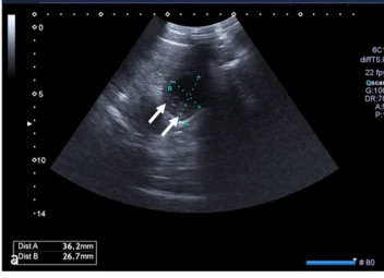
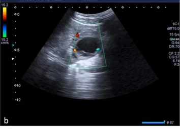
04卵巢扭轉:最不能漏的婦科急症
附件扭轉(adnexal torsion)是卵巢連同輸卵管沿血管蒂旋轉,先阻斷靜脈回流造成水腫,最後動脈也受阻而梗塞。多數發生在生育年齡,以急性或亞急性骨盆腔痛表現(Moro 2020)。診斷要靠臨床懷疑加上超音波,最後確診靠手術;就算在專科中心,術前診斷也常不準(Bar-On 2010 只有 46.1% 術前懷疑扭轉的病人在手術中證實)。
要找的徵象
- 附件變大:多數扭轉的附件明顯腫大,常合併囊腫或腫瘤當「支點」。一個 315 例的多中心研究中,扭轉器官最大徑中位數 83 mm(30–349 mm)(Moro 2020)。
- 卵巢基質水腫、濾泡被推到周邊:卵巢中央回音不均、濾泡排在皮質邊緣(Graif 1988;Moro 2020)。
- 漩渦徵象(whirlpool sign):在血管蒂上沿其長軸來回移動探頭,看到扭轉的血管蒂像漩渦般旋轉;可在灰階或彩色都卜勒下看(Vijayaraghavan 2004)。
- 都卜勒血流減少或消失、骨盆腔游離液:可以支持,但單獨的診斷力有限(見右表)。
都卜勒的侷限
- 卵巢有卵巢動脈與子宮動脈分支的雙重供血,扭轉早期或部分扭轉時動脈血流可能還在。
- 統合分析:卵巢都卜勒異常的敏感度只有 53%,特異度 95%(Garde 2023)。另一研究中異常血流的敏感度 43.8%(Bar-On 2010)。
- Moro 2020 的病例中,原始報告只有 44.2% 寫到扭轉器官沒有彩色都卜勒訊號。
- 結論:看到血流不能排除扭轉。臨床高度懷疑時,即使都卜勒正常也要照會婦產科(Mashiach 2011)。
- 在扭轉的血管蒂上同時看到動脈與靜脈血流的 5 例,術中卵巢都仍存活(Vijayaraghavan 2004,小型病例系列)。
| 超音波徵象 | 敏感度 | 特異度 | 解讀 |
|---|---|---|---|
| 卵巢水腫 | 58% | 86% | 看到有幫助,沒看到不能排除 |
| 附件腫塊 | 69% | 46% | 單獨使用診斷力差 |
| 漩渦徵象 | 65% | 91% | 特異度高,值得花時間找 |
| 卵巢都卜勒異常 | 53% | 95% | 特異度高、敏感度低 |
| 骨盆腔游離液 | 55% | 69% | 單獨使用診斷力差 |
數字取自 Garde 2023 統合分析(18 篇研究、以手術為參考標準),各分析間異質性高。兒童的統合分析中,彩色都卜勒超音波合併敏感度 78.6%、特異度 92.4%(Alsabri 2026),研究設計與成人不同,不宜直接比較。
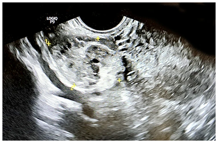
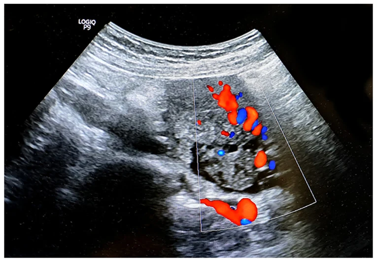
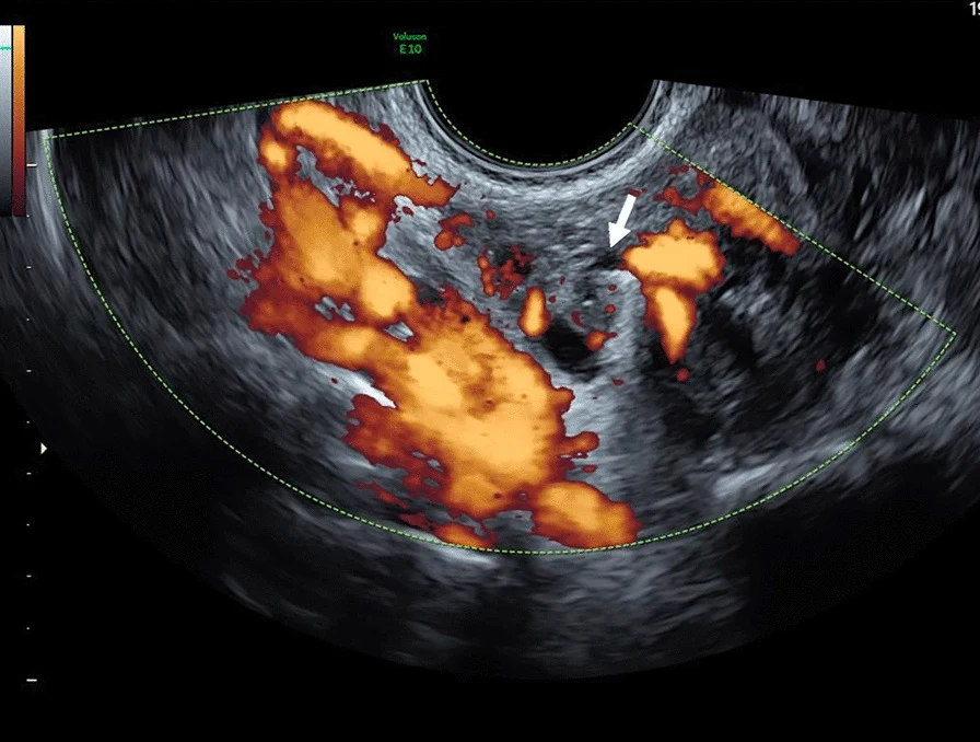
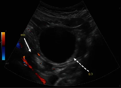
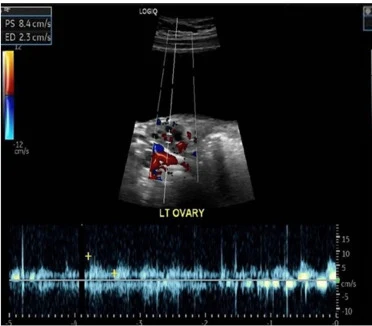
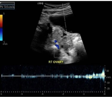
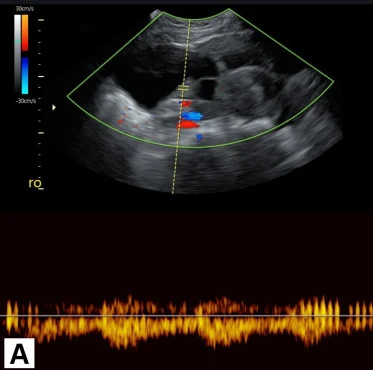
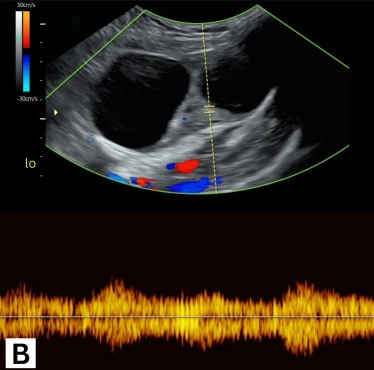
兩側卵巢都要掃,用正常側當對照比較大小、回音與血流。
以下內容需要免費訂閱
病例影像判讀與練習題開放給訂閱者。留下電子郵件即可立即閱讀全部內容;同一個瀏覽器一年內不必再輸入。換手機或電腦時,輸入同一個信箱即可。
不收費、不寄廣告。若想收到新章節通知,我們會另寄一封確認信,點了才會開始寄。詳見隱私說明。
09參考文獻與延伸閱讀
- American College of Emergency Physicians. Ultrasound guidelines: emergency, point-of-care, and clinical ultrasound guidelines in medicine. Ann Emerg Med. 2023;82(3):e115-e155. doi:10.1016/j.annemergmed.2023.06.005
- Garde I, Paredes C, Ventura L, et al. Diagnostic accuracy of ultrasound signs for detecting adnexal torsion: systematic review and meta-analysis. Ultrasound Obstet Gynecol. 2023;61(3):310-324. doi:10.1002/uog.24976
- Moro F, Bolomini G, Sibal M, et al. Imaging in gynecological disease (20): clinical and ultrasound characteristics of adnexal torsion. Ultrasound Obstet Gynecol. 2020;56(6):934-943. doi:10.1002/uog.21981
- Vijayaraghavan SB. Sonographic whirlpool sign in ovarian torsion. J Ultrasound Med. 2004;23(12):1643-1649. doi:10.7863/jum.2004.23.12.1643
- Mashiach R, Melamed N, Gilad N, Ben-Shitrit G, Meizner I. Sonographic diagnosis of ovarian torsion: accuracy and predictive factors. J Ultrasound Med. 2011;30(9):1205-1210. doi:10.7863/jum.2011.30.9.1205
- Bar-On S, Mashiach R, Stockheim D, et al. Emergency laparoscopy for suspected ovarian torsion: are we too hasty to operate? Fertil Steril. 2010;93(6):2012-2015. doi:10.1016/j.fertnstert.2008.12.022
- Graif M, Itzchak Y. Sonographic evaluation of ovarian torsion in childhood and adolescence. AJR Am J Roentgenol. 1988;150(3):647-649. doi:10.2214/ajr.150.3.647
- Alsabri M, Abady E, Elsnhory AB, Aboali AA, Rath S. Diagnostic accuracy of ultrasound for pediatric adnexal torsion: a systematic review and meta-analysis. Int J Emerg Med. 2026;19(1):90. doi:10.1186/s12245-026-01198-x
- Patel MD, Feldstein VA, Filly RA. The likelihood ratio of sonographic findings for the diagnosis of hemorrhagic ovarian cysts. J Ultrasound Med. 2005;24(5):607-614. doi:10.7863/jum.2005.24.5.607
- Levine D, Brown DL, Andreotti RF, et al. Management of asymptomatic ovarian and other adnexal cysts imaged at US: Society of Radiologists in Ultrasound consensus conference statement. Ultrasound Q. 2010;26(3):121-131. doi:10.1097/RUQ.0b013e3181f09099
- Timor-Tritsch IE, Lerner JP, Monteagudo A, Murphy KE, Heller DS. Transvaginal sonographic markers of tubal inflammatory disease. Ultrasound Obstet Gynecol. 1998;12(1):56-66. doi:10.1046/j.1469-0705.1998.12010056.x
- Lee DC, Swaminathan AK. Sensitivity of ultrasound for the diagnosis of tubo-ovarian abscess: a case report and literature review. J Emerg Med. 2011;40(2):170-175. doi:10.1016/j.jemermed.2010.02.033
- Smith-Bindman R, Kerlikowske K, Feldstein VA, et al. Endovaginal ultrasound to exclude endometrial cancer and other endometrial abnormalities. JAMA. 1998;280(17):1510-1517. doi:10.1001/jama.280.17.1510
- ACOG Committee Opinion No. 734: The role of transvaginal ultrasonography in evaluating the endometrium of women with postmenopausal bleeding. Obstet Gynecol. 2018;131(5):e124-e129. doi:10.1097/AOG.0000000000002631
- American College of Obstetricians and Gynecologists. Updated guidance regarding the role of transvaginal ultrasonography in evaluating the endometrium of individuals with postmenopausal bleeding. Obstet Gynecol. 2026;148(1):e87-e91. doi:10.1097/AOG.0000000000006275
- Boortz HE, Margolis DJ, Ragavendra N, Patel MK, Kadell BM. Migration of intrauterine devices: radiologic findings and implications for patient care. Radiographics. 2012;32(2):335-352. doi:10.1148/rg.322115068
- Di Serafino M, Iacobellis F, Schillirò ML, et al. Pelvic pain in reproductive age: US findings. Diagnostics (Basel). 2022;12(4):939. doi:10.3390/diagnostics12040939
圖片來源與授權
- 以下論文的影像在本頁裁切為單格呈現(多格拼圖拆成單格;部分影像裁去機器頂端的院所與機型字樣),圖說為本站中文改寫。部分影像以黑框遮去院名、日期與機型字樣。
- Vicente AP, Gomes AS, Jokubkiene L, et al. Ovarian borderline tumor presenting as ovarian torsion in a 17-year-old patient: a case report. J Med Case Rep 2021;15(1):7, doi:10.1186/s13256-020-02597-1(Fig. 3 a、Fig. 3 b、Fig. 2),CC BY 4.0。
- Singer SR, Melchor J, Ripps SJ, et al. Ectopic Pregnancy Observed With Kyleena Intrauterine Device Use: A Case Report. Cureus 2023;15(3):e35637, doi:10.7759/cureus.35637(Figure 1),CC BY 4.0。
- Nguyen KP, Hudspeth M, Milestone H. Spontaneous Heterotopic Pregnancy: Diagnosis and Management. Case Rep Obstet Gynecol 2022;2022:2994808, doi:10.1155/2022/2994808(Figure 4;本頁標為 Nguyen 2022a),CC BY 4.0。
- Di Serafino M, Iacobellis F, Schillirò ML, et al. Pelvic Pain in Reproductive Age: US Findings. Diagnostics (Basel) 2022;12(4):939, doi:10.3390/diagnostics12040939(Figure 3 a、Figure 3 b、Figure 4 a、Figure 4 c、Figure 6 a、Figure 13 a),CC BY 4.0。
- Varra FN, Fasoulopoulos A, Spathi E, et al. Ultrasonographic whirlpool sign in ovarian torsion with dual mature teratomas: A case report and literature review. Med Int (Lond) 2026;6(4):38, doi:10.3892/mi.2026.322(Figure 2、Figure 3),CC BY 4.0。
- Nguyen KP, Valentino WL, Bui D, et al. Ovarian Torsion: Presentation and Management in a Pediatric Patient. Case Rep Obstet Gynecol 2022;2022:9419963, doi:10.1155/2022/9419963(Figure 3;本頁標為 Nguyen 2022b),CC BY 4.0。
- Coble MS, Schoonover A, Benner C, et al. Ovarian Torsion and Its Remediation in a Three-Year-Old Girl. Cureus 2022;14(12):e32132, doi:10.7759/cureus.32132(Figure 1 left、Figure 1 right),CC BY 4.0。
- Misiriyyah N, Qurban K, Beevi A, et al. Adnexal Torsion Associated With Recurrent Ovarian Hyperstimulation Syndrome: A Case Report. Cureus 2023;15(3):e36344, doi:10.7759/cureus.36344(Figure 2 A、Figure 2 B),CC BY 4.0。
- Parchment M, Waykar M, Schellhammer S, et al. Tubo-Ovarian Abscess With Bacteremia in a Patient With an Intrauterine Contraceptive Device. Cureus 2026;18(2):e103964, doi:10.7759/cureus.103964(Figure 2、Figure 5),CC BY 4.0。
- Menendez S, Moriarty SE, Perera I, et al. Perplexing Tubo-Ovarian Abscess Presentation from an Inflammatory Process in a Patient with an Inconclusive Computed Tomography Scan. Cureus 2023;15(10):e46760, doi:10.7759/cureus.46760(Figure 2),CC BY 4.0。
- Yerrisani J, Kothari A, Collins K, et al. Evaluation of endometrial thickness by transvaginal ultrasound and baseline risk factors as a predictor for endometrial abnormalities in postmenopausal women. Australas J Ultrasound Med 2022;25(4):186-194, doi:10.1002/ajum.12311(Figure 3),CC BY 4.0。
- Koh AS. Neglected Intrauterine Device Migration Complications: Case Reports. Womens Health Rep (New Rochelle) 2023;4(1):11-18, doi:10.1089/whr.2022.0099(FIG. 5),CC BY 4.0。
- Jones DD, Kummer T, Schoen JC. Ruptured Ectopic Pregnancy with an Intrauterine Device: Case Report and Sonographic Considerations. Clin Pract Cases Emerg Med 2020;4(4):559-563, doi:10.5811/cpcem.2020.7.48258(Image 2),CC BY 4.0。