BLUE protocol:急性呼吸衰竭的肺超音波流程
01使用時機:急性呼吸衰竭
BLUE protocol(Bedside Lung Ultrasound in Emergency)是 Lichtenstein 為急性呼吸衰竭設計的床邊流程:只看肺與下肢靜脈,用固定的點組合成「profile」,指向最常見的六種病因——肺水腫、肺栓塞、肺炎、COPD、氣喘、氣胸。原作者描述整個流程不到 3 分鐘(Lichtenstein 2014)。
- 什麼時候用:急診或加護病房,病人急性喘、低血氧,主要病因還不清楚,需要在床邊先決定方向。
- 要回答的問題:這個喘比較像水(肺水腫)、發炎(肺炎)、氣道(COPD/氣喘)、血栓(肺栓塞),還是氣體(氣胸)?
- 工具:原作者強調一支微凸陣探頭、黑白機器就做得到(Lichtenstein 2015);sliding 用即時 B-mode 判斷(本課程講義)。
02六大原則:先懂肺超音波在看什麼
- 設備簡單:只調增益(gain)、深度(depth)、焦點(focus),只用 B-mode 與 M-mode,不需要彩色都卜勒。原作者用無都卜勒的灰階機器加一支微凸陣探頭,並且通常把影像濾波全部關掉(Lichtenstein 2014)。
- 氣與水交錯:胸腔內氣體與液體本來位置相反,病變讓兩者混在一起,就產生各種假影(Lichtenstein 2014)。
- 肺部徵象多半是假影(artifact):A-line、B-line 不是解剖構造,是我們拿來診斷的「訊號」。
- 所有徵象都從胸膜線出發:先找到胸膜線(bat sign),才開始判讀。
- 動態分析:肺會動,sliding、lung point、sinusoid 都要看動態,不要只看一張靜止圖。
- 急性病灶幾乎都碰到胸膜:威脅生命的急性胸腔病變大多貼著胸膜線,所以從體表就看得到(Lichtenstein 2014)。
每個假影的物理機制與完整影像,請見 肺部與胸腔超音波。
03探頭與握法
一支微凸陣探頭
- 原作者主張一支微凸陣(microconvex)探頭就能走完 BLUE:腳印小,能塞進肋間,也夠深看到後外側的實質化與積液(Lichtenstein 2014、2015)。
- 其他探頭各只提供部分資訊;太淺的線性探頭較難把 B-line 和其他彗星尾假影分開(Lichtenstein 2014;見 肺部・選探頭)。
- 設定只動增益、深度、焦點三個鈕(本課程講義);影像濾波關掉(Lichtenstein 2014)。
兩種握法(本課程講義)
- BLUE points:筆式握法。像拿筆一樣把探頭輕放在肋間,用手指微調方向。不要像握拳頭一樣用力壓——畫面不好控制,病人也不舒服。
- PLAPS point:網球拍式握法。病人仰臥時探頭要擠到後腋線甚至更背側(Lichtenstein 2014),手掌像握球拍一樣整個握住探頭,探頭垂直皮膚,再旋轉到看見清楚的 bat sign。
- 起始位置:探頭放在前胸壁,先找到清楚的 bat sign,確認看的是胸膜線,再開始判讀。
04需要的 10 個肺部徵象(複習)
BLUE 只需要十個基本徵象(Lichtenstein 2014)。這裡只列一句話重點,影像與機制請到 肺部與胸腔超音波 對應段落複習。
| 徵象 | 看到什麼 | 在 BLUE 的意義 |
|---|---|---|
| Bat sign | 兩根肋骨影夾一條胸膜線,像展翅的蝙蝠 | 起始畫面;確認找到胸膜線(正常肺) |
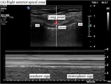
| Lung sliding/seashore sign | 胸膜線隨呼吸左右滑動;M-mode 上方水平線、下方沙灘狀 | 有 sliding=這個點排除氣胸 |
| A-line | 胸膜線下方等距重複的水平線 | 胸膜下是氣體;A-profile 的組成 |
| B-line/lung rockets | 從胸膜線出發的垂直雷射狀假影;一個肋間 3 條以上=lung rockets | 間質症候群;B-profile 的組成(B-line) |
| Quad sign | 肋骨影、胸膜線與肺線(臟層胸膜)圍出的四邊形;積液不一定無回音(膿胸、血胸可有回音) | 肋膜積液(積液) |
| Sinusoid sign | M-mode 上肺線吸氣時往胸膜線靠近,畫出正弦波 | 肋膜積液,且黏稠度低 |
| Tissue-like sign | 肺變得像肝臟一樣的實質回音(hepatization) | 大片實質化(實質化) |
| Shred(fractal)sign | 實質化與含氣肺之間鋸齒狀、碎裂的邊界 | 實質化(未及整葉) |
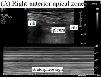
| Stratosphere sign | M-mode 全部是水平線(barcode),沒有沙灘 | sliding 消失;可能是氣胸(氣胸) |
| Lung point | sliding 與無 sliding 在同一點交替出現 | 氣胸的確診徵象 |
十個徵象依 Lichtenstein 2014、2015 的清單。講義另外補充的 O-line(既沒有 A-line 也沒有 B-line)、C-line(前胸胸膜下的小低回音實質化)、air/fluid bronchogram 屬延伸觀念,見下方。
四個「幾乎一定」:④長,一路延伸到螢幕底部不衰減 ⑤邊界清楚、像雷射 ⑥高回音 ⑦會抹掉 A-line(Lichtenstein 2014)。這個定義把 B-line 和 Z-line、E-line 等其他垂直假影分開。
Lung rockets 代表肺泡-間質症候群:放射學上瀰漫肺泡-間質症候群的病人,93.4% 全肺表面都有多條 comet-tail;胸部 X 光正常者 93.0% 沒有(或只限於最外側肋間)(Lichtenstein 1997)。前胸以 A-line 為主時,預測肺動脈楔壓(PAOP)≦18 mmHg 的特異度 93%;但 B-line 為主可見於很寬的 PAOP 範圍,不能反推壓力(Lichtenstein 2009b)。原作者在 FALLS protocol 把 A-line 轉成 lung rockets 的門檻放在 PAOP 18 mmHg 左右(Lichtenstein 2015)——這是「由乾轉濕」的轉折點,不代表看到 B-line 就能說壓力一定高於 18。
- Lung point:66 例氣胸中 44 例找得到(敏感度 66%),對照組 0 例(特異度 100%)(Lichtenstein 2000)。找到就確診,找不到不能排除。
- 有 lung sliding:可以立即排除該處的前胸氣胸;原始研究中 sliding 消失對氣胸的敏感度 95.3%、特異度 91.1%(Lichtenstein 1995)。
- Dynamic air bronchogram:實質化裡的亮點隨呼吸往外移動。在有 air bronchogram 的實質化中,對肺炎(相對於吸收性肺塌陷)特異度 94%、敏感度 61%;static air bronchogram 見於多數肺塌陷,也見於約三分之一的肺炎(Lichtenstein 2009a)。
- Fluid bronchogram:實質化裡管狀無回音的支氣管,代表支氣管內是液體(本課程講義)。
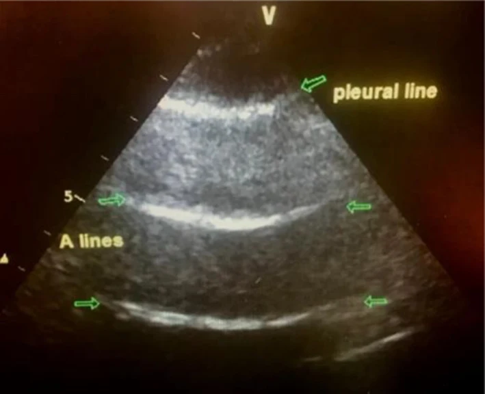
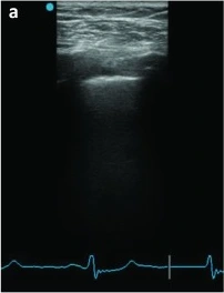
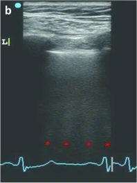
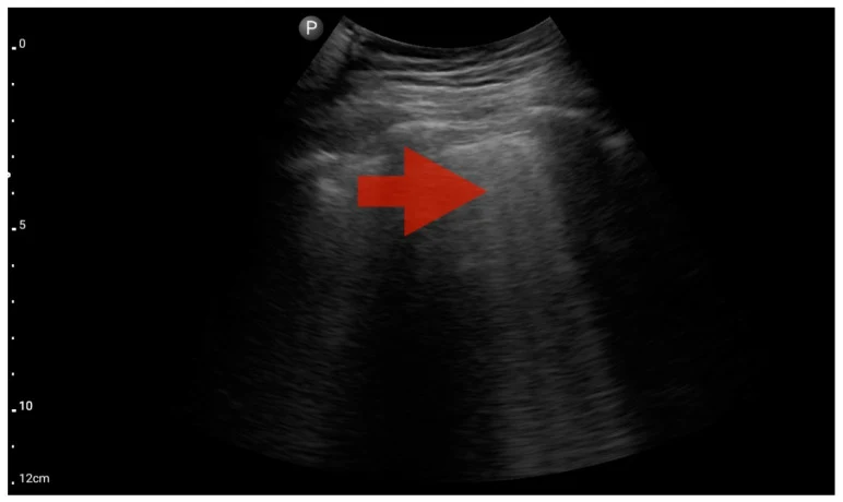
Haaksma 2019 這個病例是肺出血造成 A/B-profile,提醒我們 profile 是「指向」而不是診斷本身。
05三個掃描點與 PLAPS 的 CT 觀念
- 雙手定位:兩手大小比照病人的手,上手碰鎖骨、拇指不算,兩手並排就蓋住一側前胸(Lichtenstein 2014)。
- Upper BLUE point:上手第三、四指根之間(本課程講義;Lichtenstein 2014 寫作「上手中央」,兩者相同位置)。問:有沒有氣胸?有沒有間質症候群?
- Lower BLUE point:下手掌心中央。同樣問氣胸、間質症候群,順便注意積液。
- PLAPS point:Lower BLUE point 的水平線往後延伸,與後腋線(posterior axillary line)的交點。問:有沒有實質化或積液(posterolateral alveolar and/or pleural syndrome)?
06Profile 定義與決策流程
| Profile | 定義(前胸 BLUE points) |
|---|---|
| A-profile | lung sliding+A-line(雙側) |
| A′-profile | A-line,但 sliding 消失 → 去找 lung point |
| Pseudo-A′-profile | A-line、sliding 消失,但看得到 lung pulse(M-mode 上即 T line)(本課程講義);表示兩層胸膜仍相貼,sliding 消失另有原因,例如沾黏 |
| B-profile | lung sliding+lung rockets(雙側瀰漫 B-line) |
| B′-profile | lung rockets,但 sliding 消失 |
| A/B-profile | 一側 A-profile、另一側 B-profile |
| C-profile | 前胸實質化,不論大小與數量;增厚不規則的胸膜線視同(Lichtenstein 2014) |
| Nude profile | A-profile、無 DVT、無 PLAPS(A-no-V-no-PLAPS) |
定義依 Lichtenstein 2014;pseudo-A′ 為本課程講義補充。
肺水腫的漏出液像「油」,sliding 保留(B-profile);肺炎的滲出液像「膠」,sliding 消失(B′-profile)——這是原作者對兩者差異的解釋(Lichtenstein 2014)。
完整決策流程
- 掃兩側 Upper/Lower BLUE point:sliding 有沒有?A 還是 B?有沒有實質化?
- B-profile(且沒有 C-line、沒有 shred sign)→ 肺水腫。
- B′、A/B、C-profile,或任何 BLUE point 出現 C-line、shred sign → 肺炎(或 ARDS)(本課程講義)。
- A-profile → 下肢靜脈:有 DVT → 肺栓塞。
- A-profile、無 DVT → PLAPS point:有 PLAPS → 肺炎;沒有(nude profile)→ COPD 或氣喘。
- A′-profile → 找 lung point:有 → 氣胸;沒有 → 需要其他影像檢查。
| 八種組合 | 指向 |
|---|---|
| ① A-profile+DVT | 肺栓塞 |
| ② A-no-V-PLAPS | 肺炎 |
| ③ A-no-V-no-PLAPS(nude) | COPD/氣喘 |
| ④ B-profile | 心因性肺水腫 |
| ⑤ B′-profile | 肺炎 |
| ⑥ A/B-profile | 肺炎 |
| ⑦ C-profile | 肺炎 |
| ⑧ A′-profile+lung point | 氣胸 |
依 Lichtenstein 2008 決策樹整理。決策樹的目標是快速分流、整體正確率約 90.5%,不是 100%。
以下內容需要免費訂閱
病例影像判讀與練習題開放給訂閱者。留下電子郵件即可立即閱讀全部內容;同一個瀏覽器一年內不必再輸入。換手機或電腦時,輸入同一個信箱即可。
不收費、不寄廣告。若想收到新章節通知,我們會另寄一封確認信,點了才會開始寄。詳見隱私說明。
14參考文獻與延伸閱讀
- Lichtenstein DA, Mezière GA. Relevance of lung ultrasound in the diagnosis of acute respiratory failure: the BLUE protocol. Chest. 2008;134(1):117-125. doi:10.1378/chest.07-2800
- Lichtenstein DA. BLUE-protocol and FALLS-protocol: two applications of lung ultrasound in the critically ill. Chest. 2015;147(6):1659-1670. doi:10.1378/chest.14-1313
- Lichtenstein DA. Lung ultrasound in the critically ill. Ann Intensive Care. 2014;4(1):1. doi:10.1186/2110-5820-4-1
- Lichtenstein D, Mézière G, Biderman P, Gepner A, Barré O. The comet-tail artifact. An ultrasound sign of alveolar-interstitial syndrome. Am J Respir Crit Care Med. 1997;156(5):1640-1646. doi:10.1164/ajrccm.156.5.96-07096
- Lichtenstein DA, Menu Y. A bedside ultrasound sign ruling out pneumothorax in the critically ill. Lung sliding. Chest. 1995;108(5):1345-1348. doi:10.1378/chest.108.5.1345
- Lichtenstein D, Mezière G, Biderman P, Gepner A. The "lung point": an ultrasound sign specific to pneumothorax. Intensive Care Med. 2000;26(10):1434-1440. doi:10.1007/s001340000627
- Lichtenstein D, Mezière G, Seitz J. The dynamic air bronchogram. A lung ultrasound sign of alveolar consolidation ruling out atelectasis. Chest. 2009;135(6):1421-1425.(文中 2009a) doi:10.1378/chest.08-2281
- Lichtenstein DA, Mezière GA, Lagoueyte JF, Biderman P, Goldstein I, Gepner A. A-lines and B-lines: lung ultrasound as a bedside tool for predicting pulmonary artery occlusion pressure in the critically ill. Chest. 2009;136(4):1014-1020.(文中 2009b) doi:10.1378/chest.09-0001
- Ding X, Wang X, Chen H, Zhao H, Chao Y, Yin W, Liu D; Chinese Critical Ultrasound Study Group. [A comparative study of different bedside lung ultrasound examination for the location and signs of diaphragmatic points]. Zhonghua Nei Ke Za Zhi. 2015;54(9):778-782.(中文)
- Bekgoz B, Kilicaslan I, Bildik F, Keles A, et al. BLUE protocol ultrasonography in Emergency Department patients presenting with acute dyspnea. Am J Emerg Med. 2019;37(11):2020-2027. doi:10.1016/j.ajem.2019.02.028
- Chaitra S, Hattiholi VV. Diagnostic accuracy of Bedside Lung Ultrasound in Emergency protocol for the diagnosis of acute respiratory failure. J Med Ultrasound. 2022;30(2):94-100. doi:10.4103/JMU.JMU_25_21
- Buessler A, Chouihed T, Duarte K, Bassand A, et al. Accuracy of several lung ultrasound methods for the diagnosis of acute heart failure in the ED: a multicenter prospective study. Chest. 2020;157(1):99-110. doi:10.1016/j.chest.2019.07.017
- Zanobetti M, Scorpiniti M, Gigli C, Nazerian P, et al. Point-of-care ultrasonography for evaluation of acute dyspnea in the ED. Chest. 2017;151(6):1295-1301. doi:10.1016/j.chest.2017.02.003
- Silva S, Biendel C, Ruiz J, Olivier M, et al. Usefulness of cardiothoracic chest ultrasound in the management of acute respiratory failure in critical care practice. Chest. 2013;144(3):859-865. doi:10.1378/chest.13-0167
- Laursen CB, Sloth E, Lassen AT, Christensen Rd, et al. Point-of-care ultrasonography in patients admitted with respiratory symptoms: a single-blind, randomised controlled trial. Lancet Respir Med. 2014;2(8):638-646. doi:10.1016/S2213-2600(14)70135-3
- Zaalouk TM, Bitar ZI, Maadarani OS, Ragab Elshabasy RD. Modified BLUE protocol ultrasonography can diagnose thrombotic complications of COVID-19 with normal lung ultrasound. Clin Case Rep. 2021;9(5):e04075. doi:10.1002/ccr3.4075
- Nazerian P, Vanni S, Volpicelli G, Gigli C, et al. Accuracy of point-of-care multiorgan ultrasonography for the diagnosis of pulmonary embolism. Chest. 2014;145(5):950-957. doi:10.1378/chest.13-1087
圖片來源與授權
- 以下論文的多格拼圖在本頁拆成單格裁切呈現,圖說為本站中文改寫。
- Haaksma ME, Nossent EJ, Elbers P, Tuinman PR. Case Report: Pulmonary hemorrhage as a rare cause of lung ultrasound A/B-profile. F1000Res 2019;8:788, doi:10.12688/f1000research.19329.2(Fig. 1a、1b),CC BY 4.0。
- Zaalouk TM, Bitar ZI, Maadarani OS, Ragab Elshabasy RD. Modified BLUE protocol ultrasonography can diagnose thrombotic complications of COVID-19 with normal lung ultrasound. Clin Case Rep 2021;9(5):e04075, doi:10.1002/ccr3.4075(Fig. 2、3、4),CC BY 4.0。
- Kowalczyk D, Turkowiak M, Piotrowski WJ, Rosiak O, Białas AJ. Unlocking diagnostic precision: FATE protocol integration with BLUE and eFAST protocols for enhanced pre-hospital differential diagnosis of pleural effusion manifested as dyspnea in adults—a pilot study. J Clin Med 2024;13(6):1573, doi:10.3390/jcm13061573(Fig. 8、9、11、12、13、14、15),CC BY 4.0。
- Chae MS, Kwak J, Roh K, Kim M, et al. Pneumoperitoneum-induced pneumothorax during laparoscopic living donor hepatectomy: a case report. BMC Surg 2020;20:206, doi:10.1186/s12893-020-00868-8(Fig. 1a、2a),CC BY 4.0。
- Khalid WKK, Ibrahim HAM, Al Hoqani IAA. Fast breathing, faster thinking: how the Bedside Lung Ultrasound in Emergency (BLUE) protocol prevented a missed pulmonary embolism. Cureus 2026;18(5):e108755, doi:10.7759/cureus.108755(Fig. 1A、1B),CC BY 4.0。
- 決策樹、雙手定位與 PLAPS 橫切面示意為本站依 Lichtenstein 2008、2014 原文與本課程講義重繪的示意圖。