超音波圖譜 › 第 11 章 · 主動脈
主動脈超音波
這一章的重點 腹主動脈從橫膈一路掃到分叉,短軸外壁到外壁量,≥ 3 cm 就是動脈瘤;超音波看得出「有沒有瘤」,但看不出「有沒有破」。主動脈剝離則用心臟切面看根部是否擴張、有沒有內膜瓣與心包膜積液:看到直接徵象很有用,看不到不能排除。
01使用時機:哪些病人要掃主動脈
主動脈急症(腹主動脈瘤破裂、急性主動脈症候群)常以腹痛、背痛、胸痛、暈厥或休克來表現,而且典型症狀常常不齊全。床邊超音波的角色是快速回答兩個問題:有沒有動脈瘤?有沒有剝離的徵象?然後決定誰要立刻 CT、會診或轉送。
學完這一章,你應該能夠
- 從橫膈到分叉完整掃過腹主動脈,並在短軸、外壁到外壁量出最大外徑。
- 說出超音波為什麼排除不了 AAA 破裂,以及看到瘤+不穩定時該怎麼做。
- 在胸骨旁長軸找出主動脈根部與降主動脈,認出內膜瓣、根部擴張、心包膜積液。
- 避開 IVC 誤認、斜切高估、橫膈腳偽內膜瓣等陷阱。
什麼時候想到要掃
- 50 歲以上的腹痛、腰背痛、像腎絞痛的疼痛(AAA 要放進鑑別診斷)。
- 不明原因的低血壓或暈厥:放進休克評估流程一起看,見 休克與 RUSH。
- 撕裂樣胸背痛、兩側脈搏或血壓不對稱、合併神經學症狀的胸痛。
- 胸痛合併心包膜積液、新出現的主動脈瓣逆流,或腹痛合併下肢缺血。
Rubano 2013 統合 7 篇研究:症狀病人中,約一半的 AAA 病人沒有低血壓、背痛、腹部搏動性腫塊這組典型三聯徵。所以不要等「教科書表現」才拿探頭。
02解剖:分段、分支與 IVC 的區別
腹主動脈三段(本課程講義)
- 近端:橫膈到腹腔幹(celiac trunk)。
- 中段:腹腔幹到上腸繫膜動脈(SMA),左右腎動脈在 SMA 下方不遠處分出。
- 遠端:腎動脈以下到分叉(約臍部高度)成為左右總髂動脈。大多數 AAA 位在腎動脈以下,所以遠端一定要掃到。
短軸的定位地標
- 脊椎體前方、偏病人左側的圓形搏動血管是主動脈;偏右的是 IVC。
- 腹腔幹向前分出後分成肝動脈與脾動脈(「海鷗」形狀)。
- SMA 在其下方向前分出,前方有脾靜脈橫過;左腎靜脈夾在 SMA 與主動脈之間進入 IVC。
- 看到這三個地標,就知道自己在哪一段。
| 主動脈 | 下腔靜脈(IVC) | |
|---|---|---|
| 位置 | 脊椎前方偏左 | 脊椎前方偏右,穿過肝臟後方 |
| 管壁 | 厚、高回音,可有鈣化 | 薄 |
| 搏動 | 明顯,與心跳一致 | 隨呼吸改變管徑,可有雙相波動 |
| 探頭加壓 | 不易壓扁 | 可被壓扁 |
| 分支 | 腹腔幹、SMA 向前分出 | 肝靜脈在上端匯入 |
本表為講義重點。單看搏動不可靠:IVC 會傳導主動脈的搏動,主動脈內有大量血栓時搏動也可能不明顯;不確定時用彩色都卜勒或波形確認,並找分支地標。
03腹主動脈掃描技巧
探頭與姿勢
- 凸面探頭(講義 2.5–5 MHz),腹部預設;需要時用相位陣列(心臟)探頭從肋下切入。
- 病人平躺;屈髖屈膝讓腹壁放鬆(講義)。
- 橫切時探頭標記朝病人右側,縱切時朝頭側,配合螢幕標記,維持一致方向。
- 深度先開深一點找到脊椎,再把主動脈拉到畫面中央。
腸氣擋住時(講義)
- 溫和、穩定、持續地往下壓,把腸氣推開;不要用力猛壓(有瘤的病人要溫柔)。
- 換左側臥位,從右側腹部以肝臟當窗口往下看。
- 從左側腰部冠狀切面(經腰窗口,translumbar)看主動脈與 IVC。
- 講義也提到讓病人喝水充滿胃部作為聲窗。
- 劍突下橫切找到脊椎與主動脈,確認近端(腹腔幹)。
- 往尾側滑動,一路追到 SMA、腎動脈,再到分叉;每一段都要看到完整圓形管壁。
- 在分叉處看左右總髂動脈的起始段。
- 轉縱切補看全長,確認沒有局部膨大或內膜瓣。
- 在最粗處量外徑(見下一節);看不清楚的段落要寫明「未完整評估」。
沒有掃到分叉,就不算陰性 AAA 多在腎動脈以下。只在上腹看到一段正常主動脈就說「沒有 AAA」,是常見而危險的錯誤;看不到的段落要說出來,必要時安排 CT。
04量測:短軸、外壁到外壁
怎麼量
- 在短軸、與血管長軸垂直的切面量。
- 外壁到外壁(outer wall to outer wall),把壁上血栓一起算進去;只量有血流的管腔會低估。
- 前後徑與左右徑都量,取最大值。
- 主動脈彎曲時橫切容易斜切成橢圓,長徑會高估;轉探頭找到最圓的切面,或改用縱切確認。
幾公分算異常
| 部位 | 數值 | 意義 |
|---|---|---|
| 腹主動脈 | ≥ 3 cm | AAA 定義(Rubano 2013 等急診研究採用) |
| 胸主動脈(根部/升主動脈) | ≥ 4 cm | 急診 FOCUS 研究常用的擴張切點(Taylor 2012) |
動脈瘤大小與追蹤時程由血管外科處理(Chaikof 2018)。急診的任務是:有沒有瘤、病人穩不穩、要不要馬上叫人。
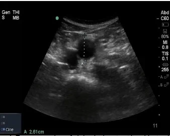
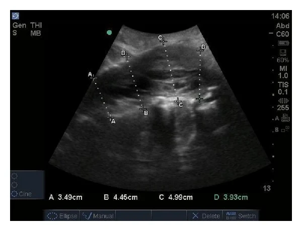
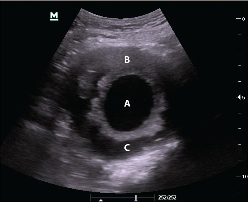
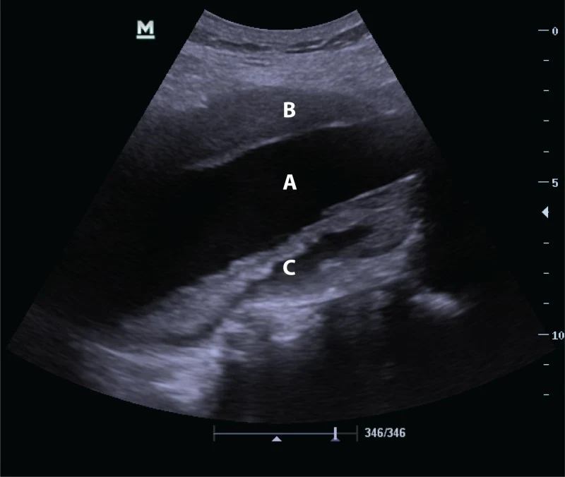
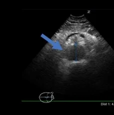
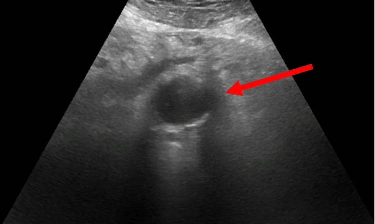
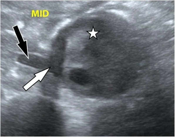
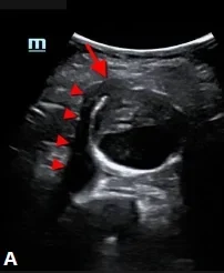
影像來源:Earl-Royal 2019、Tsung 2013、Goldstein 2019、Yee 2017、Huang 2022、Diaz 2020、Sherman 2026,裁切單格,CC BY。下排兩張是少數「看得到」破裂徵象的病例,不代表破裂通常看得到(見下一節)。
05破裂:看得到瘤,常常看不到破
證據
- 找瘤很準:急診醫師床邊超音波診斷 AAA,統合敏感度 99%、特異度 98%(Rubano 2013,7 篇研究、655 位病人);單中心前瞻研究敏感度 100%、特異度 98%(Tayal 2003)。
- 找破裂很差:疑似 AAA 滲漏的 60 位病人,超音波判斷有無動脈瘤正確率 98%,但偵測主動脈外出血的敏感度只有 4%(Shuman 1988)。
- 原因:破裂多出血到後腹腔,被腸氣與深度擋住,不一定流進腹腔,FAST 也常是陰性(本課程講義)。
臨床決策
- 看到 AAA+症狀+不穩定:視為破裂,立刻聯絡血管外科/手術室,不要為了 CT 拖延。Shuman 1988 中,「有瘤+腹痛+血行動力學不穩定」讓 22 位中 21 位做出正確的緊急手術決定。
- 看到 AAA+症狀+穩定:儘快 CTA,同時通知血管外科。
- 看不到 AAA 且全段掃完:AAA 破裂的可能性大幅降低,回頭找其他原因。
- 沒掃完整:當作沒做,安排其他影像。
腹腔沒有積液,不能排除 AAA 破裂 後腹腔出血用超音波很難看到。有症狀的病人只要看到動脈瘤,就要當作可能破裂處理;SVS 指引建議破裂 AAA 從到院到介入在 90 分鐘以內(Chaikof 2018)。
以下內容需要免費訂閱
病例影像判讀與練習題開放給訂閱者。留下電子郵件即可立即閱讀全部內容;同一個瀏覽器一年內不必再輸入。換手機或電腦時,輸入同一個信箱即可。
不收費、不寄廣告。若想收到新章節通知,我們會另寄一封確認信,點了才會開始寄。詳見隱私說明。
10參考文獻與延伸閱讀
- Rubano E, Mehta N, Caputo W, Paladino L, Sinert R. Systematic review: emergency department bedside ultrasonography for diagnosing suspected abdominal aortic aneurysm. Acad Emerg Med. 2013;20(2):128-138. doi:10.1111/acem.12080
- Tayal VS, Graf CD, Gibbs MA. Prospective study of accuracy and outcome of emergency ultrasound for abdominal aortic aneurysm over two years. Acad Emerg Med. 2003;10(8):867-871. doi:10.1111/j.1553-2712.2003.tb00630.x
- Shuman WP, Hastrup W Jr, Kohler TR, et al. Suspected leaking abdominal aortic aneurysm: use of sonography in the emergency room. Radiology. 1988;168(1):117-119. doi:10.1148/radiology.168.1.3289085
- Chaikof EL, Dalman RL, Eskandari MK, et al. The Society for Vascular Surgery practice guidelines on the care of patients with an abdominal aortic aneurysm. J Vasc Surg. 2018;67(1):2-77.e2. doi:10.1016/j.jvs.2017.10.044
- Isselbacher EM, Preventza O, Hamilton Black J 3rd, et al. 2022 ACC/AHA guideline for the diagnosis and management of aortic disease. Circulation. 2022;146(24):e334-e482. doi:10.1161/CIR.0000000000001106
- Goldstein SA, Evangelista A, Abbara S, et al. Multimodality imaging of diseases of the thoracic aorta in adults: from the American Society of Echocardiography and the European Association of Cardiovascular Imaging. J Am Soc Echocardiogr. 2015;28(2):119-182. doi:10.1016/j.echo.2014.11.015
- Taylor RA, Oliva I, Van Tonder R, Elefteriades J, Dziura J, Moore CL. Point-of-care focused cardiac ultrasound for the assessment of thoracic aortic dimensions, dilation, and aneurysmal disease. Acad Emerg Med. 2012;19(2):244-247. doi:10.1111/j.1553-2712.2011.01279.x
- Nazerian P, Vanni S, Castelli M, et al. Diagnostic performance of emergency transthoracic focus cardiac ultrasound in suspected acute type A aortic dissection. Intern Emerg Med. 2014;9(6):665-670. doi:10.1007/s11739-014-1080-9
- Nazerian P, Mueller C, Vanni S, et al. Integration of transthoracic focused cardiac ultrasound in the diagnostic algorithm for suspected acute aortic syndromes. Eur Heart J. 2019;40(24):1952-1960. doi:10.1093/eurheartj/ehz207
- Pare JR, Liu R, Moore CL, et al. Emergency physician focused cardiac ultrasound improves diagnosis of ascending aortic dissection. Am J Emerg Med. 2016;34(3):486-492. doi:10.1016/j.ajem.2015.12.005
- Haaz WS, Mintz GS, Kotler MN, Parry W, Segal BL. Two dimensional echocardiographic recognition of the descending thoracic aorta: value in differentiating pericardial from pleural effusions. Am J Cardiol. 1980;46(5):739-743. doi:10.1016/0002-9149(80)90423-3
圖片來源與授權
- 以下論文的影像在本頁裁切為單格呈現(多格拼圖拆成單格;部分影像裁去機型字樣或以黑框遮蓋日期時間),圖說為本站中文改寫。
- Earl-Royal E, Nguyen PD, Alvarez A, et al. Detection of Type B Aortic Dissection in the Emergency Department with Point-of-Care Ultrasound. Clin Pract Cases Emerg Med 2019;3(3):202-207, doi:10.5811/cpcem.2019.5.42928(Image 1、3),CC BY 4.0。
- Tsung AH, Nickels LC, De Portu G, et al. Aortic dissection and thrombosis diagnosed by emergency ultrasound in a patient with leg pain and paralysis. Case Rep Vasc Med 2013;2013:490126, doi:10.1155/2013/490126(Fig. 2),CC BY 3.0。
- Goldstein LN, Wells M. Point-of-care Ultrasound Trumps Computed Tomography in a Case of Abdominal Aortic Aneurysm Assessment. Cureus 2019;11(10):e5989, doi:10.7759/cureus.5989(Fig. 1),CC BY 3.0。
- Yee AM, Etebari CV, Adhikari S, et al. Point of Care Ultrasound Diagnosis of a Massive Thoracoabdominal Aortic Aneurysm. Cureus 2017;9(8):e1611, doi:10.7759/cureus.1611(Fig. 1A、2A),CC BY 3.0。
- Diaz O, Eilbert W. Ruptured abdominal aortic aneurysm identified on point-of-care ultrasound in the emergency department. Int J Emerg Med 2020;13(1):25, doi:10.1186/s12245-020-00279-9(Fig. 1),CC BY 4.0。
- Sherman MH. Ruptured Abdominal Aortic Aneurysm Identified on Point-of-Care Ultrasound. Clin Pract Cases Emerg Med 2026;10(3):424-425, doi:10.5811/cpcem.61897(Image A),CC BY 4.0。
- Huang D, Huttleston A, Fraunfelter F, et al. Penetrating Aortic Ulceration With Pseudoaneurysm and Intramural Hematoma: Emergency Department Management and Point-of-Care Ultrasound Diagnosis. Cureus 2022;14(7):e27536, doi:10.7759/cureus.27536(Fig. 1),CC BY 3.0。
- Craen A, Rosario J, Amico K, et al. Transthoracic Echocardiographic Findings of Stanford Type A Aortic Dissection: A Case Report. Cureus 2019;11(11):e6207, doi:10.7759/cureus.6207(Fig. 1、3、4),CC BY 3.0。
- Thomas K, Amr O. Use of Point-of-Care Ultrasound for Early Identification of Acute Aortic Root Dissection. Case Rep Crit Care 2022;2022:7166230, doi:10.1155/2022/7166230(Fig. 3、4),CC BY 4.0。
- Londono S, Mendoza-Garcia FC, Kohen B. Case of Aortic Dissection Diagnosed by Suprasternal Notch Point-of-Care Ultrasound (POCUS) in a 73-Year-Old Man With Chest and Lower Extremity Pain. Cureus 2026;18(6):e110529, doi:10.7759/cureus.110529(Fig. 1),CC BY 4.0。
- Zarama V, Arango-Granados MC, Bustamante Cristancho LA. Importance of Multiple-window Assessment for the Diagnosis of Ascending Aortic Dissection Using Point-of-care Ultrasound: Report of Three Cases. Clin Pract Cases Emerg Med 2019;3(4):333-337, doi:10.5811/cpcem.2019.6.43245(Image 1、3),CC BY 4.0。
- Abrams E, Allen A, Lahham S. Aortic Dissection with Subsequent Hemorrhagic Tamponade Diagnosed with Point-of-care Ultrasound in a Patient Presenting with STEMI. Clin Pract Cases Emerg Med 2019;3(2):103-106, doi:10.5811/cpcem.2019.1.40869(Image 2、3),CC BY 4.0。
- Langberg S, Favot M. Hiatal Hernia Mimicking Aortic Aneurysm on Point-of-care Echocardiography. Clin Pract Cases Emerg Med 2017;1(4):419-420, doi:10.5811/cpcem.2017.6.33450(Image),CC BY 4.0。
- Klee O, Buechler J, Fears M, et al. Erratum: Normal Anatomy Mimicking an Abdominal Aortic Dissection. POCUS J 2026;11(1):50-53, doi:10.24908/pocusj.v11i01.20821(Fig. 1、5),CC BY 4.0。
- Cárdenas López V, Blanco P. Acute Type A Aortic Dissection Diagnosed by POCUS in a 29-year-old Man. POCUS J 2023;8(2):118-120, doi:10.24908/pocus.v8i2.16533(Fig. 1A),CC BY 4.0。
- Wroblewski R, Gibbons R, Costantino T. Point-of-care Ultrasound Diagnosis of an Atypical Acute Aortic Dissection. Clin Pract Cases Emerg Med 2018;2(4):300-303, doi:10.5811/cpcem.2018.6.38106(Image 1),CC BY 4.0。